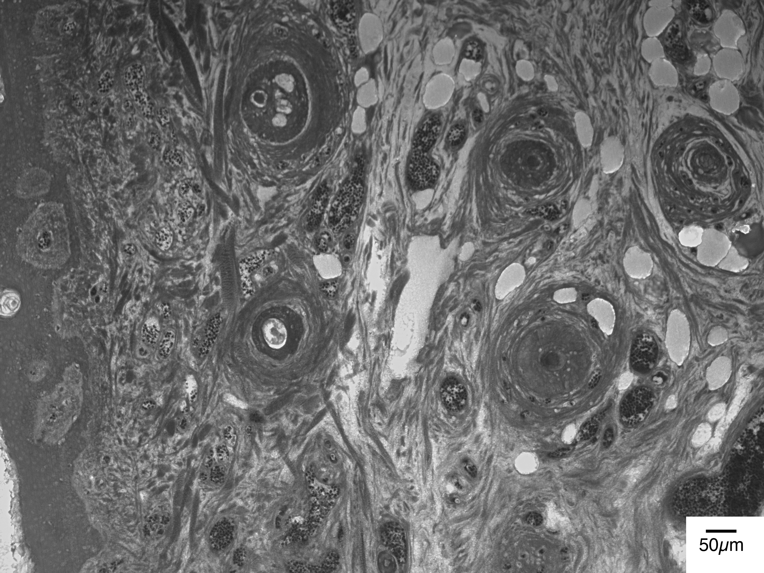
| Human
(Homo sapiens sapiens) Adult skin |
Movie Grey-scale (jpg file) |
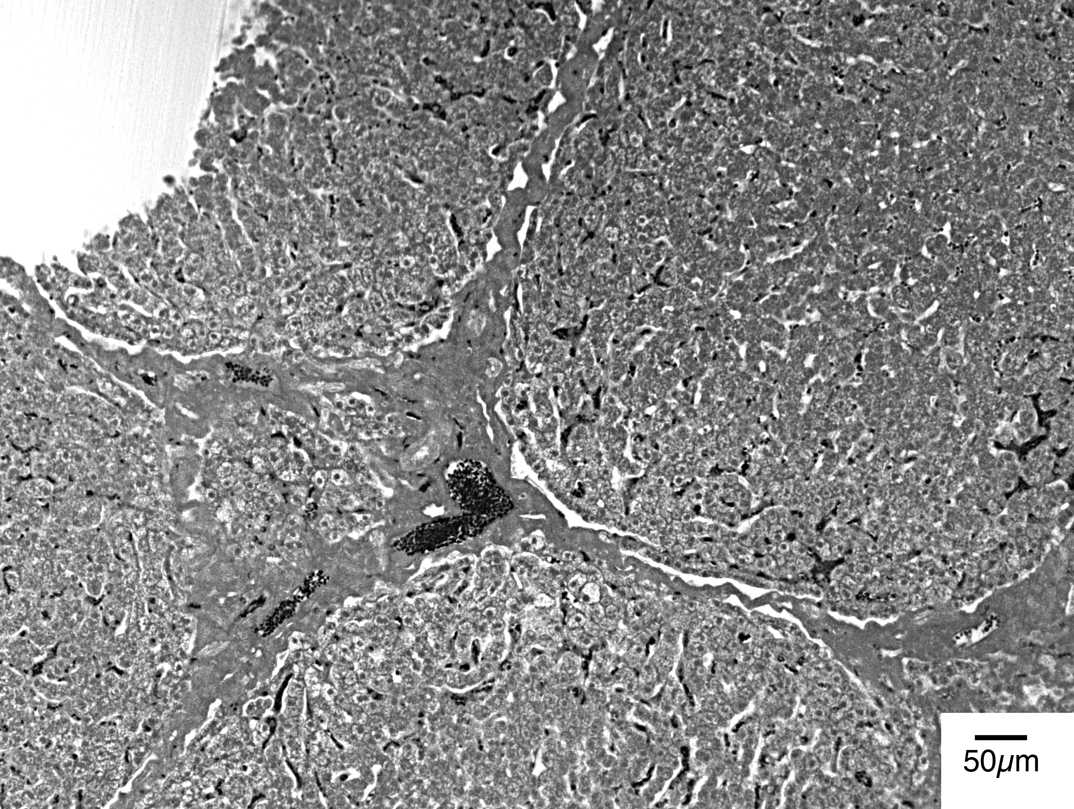
| Pig
(Sus scrofa domesticus) Adult liver |
Movie 1 Grey-scale 1 (jpg file) Movie 2 Grey-scale 2 (jpg file) |
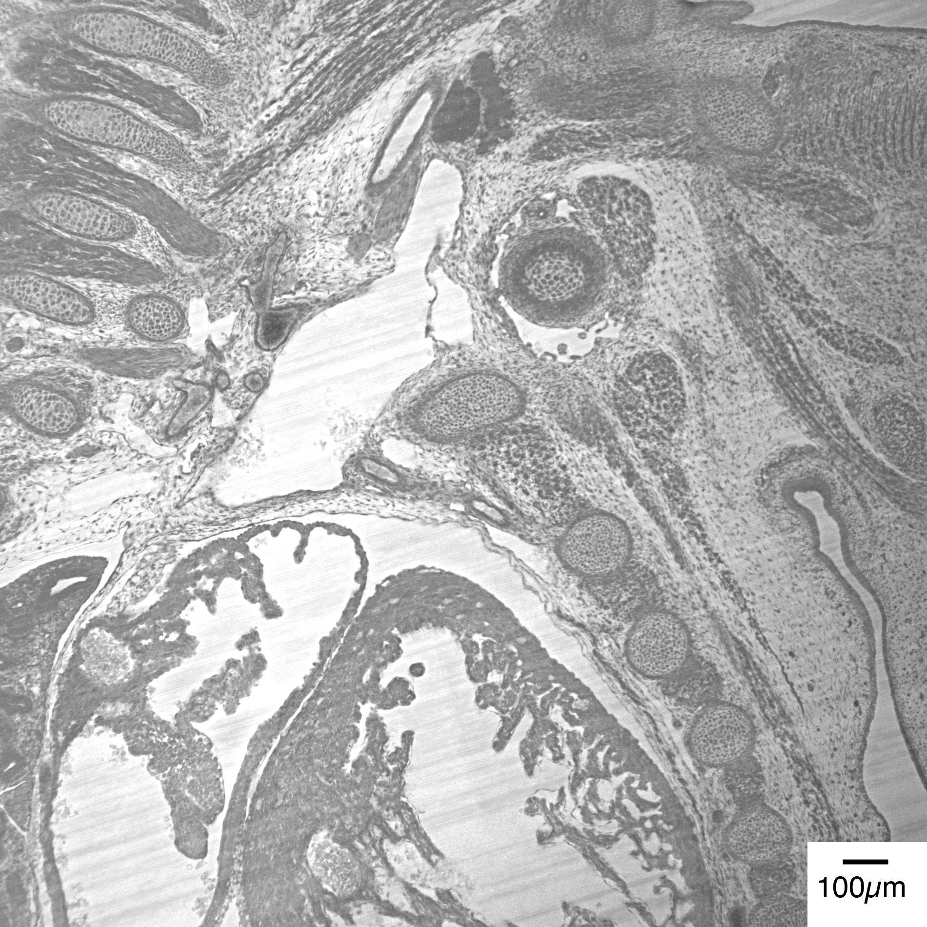
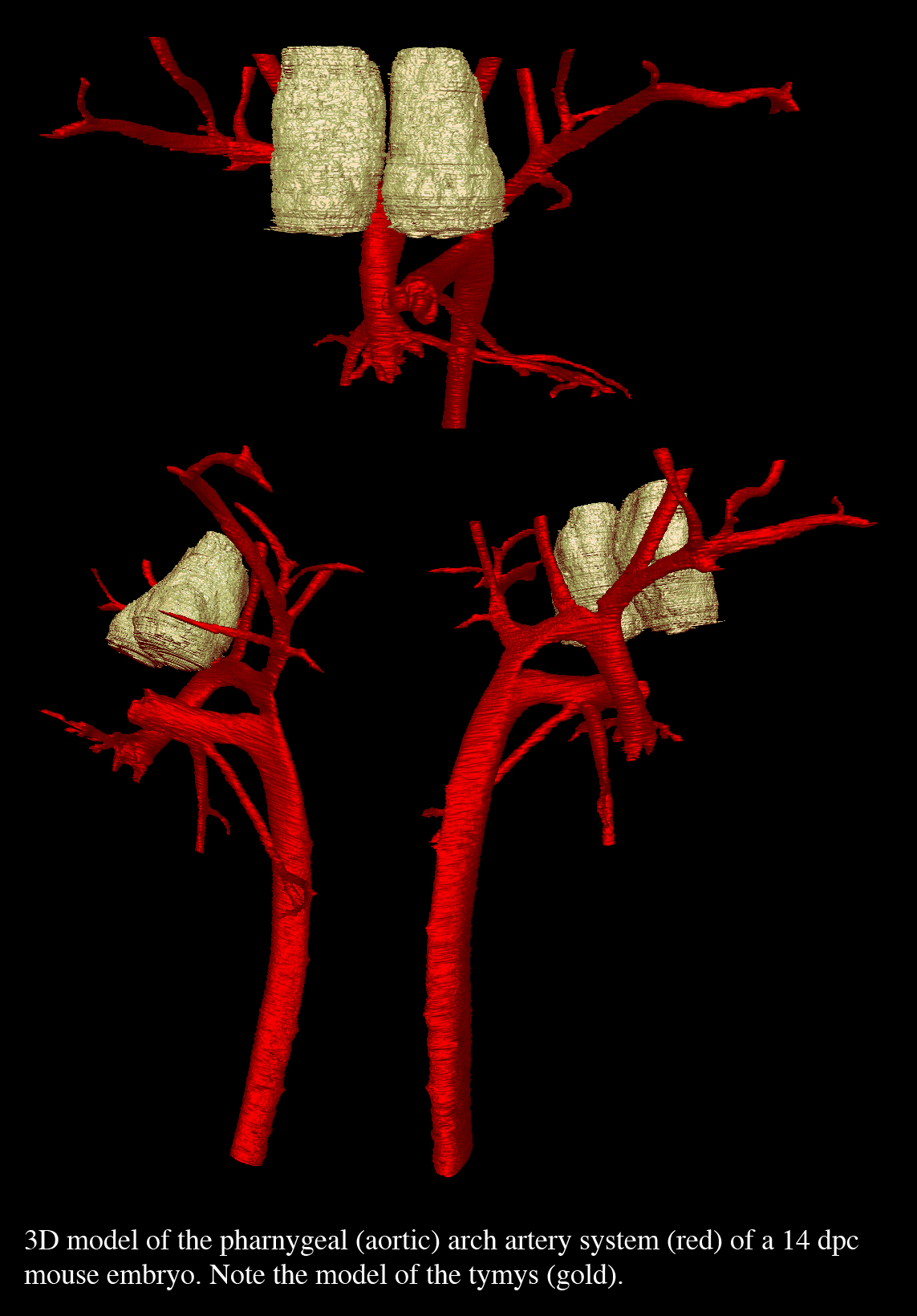
| Mouse
(Mus musculus) Thorax of an embryos of 14.5 dpc |
Movie Grey-scale (jpg file) Colour jpg of 3D model |
| Chick
(Gallus domesticus) Thorax of an embryo of HH 34 |
Movie Grey-scale (jpg file) Colour jpg of 3D model |
| Quail
(Coturnix
japonica) Thorax of an embryo corresponding to HH 20 |
Movie Grey-scale (jpg file) |
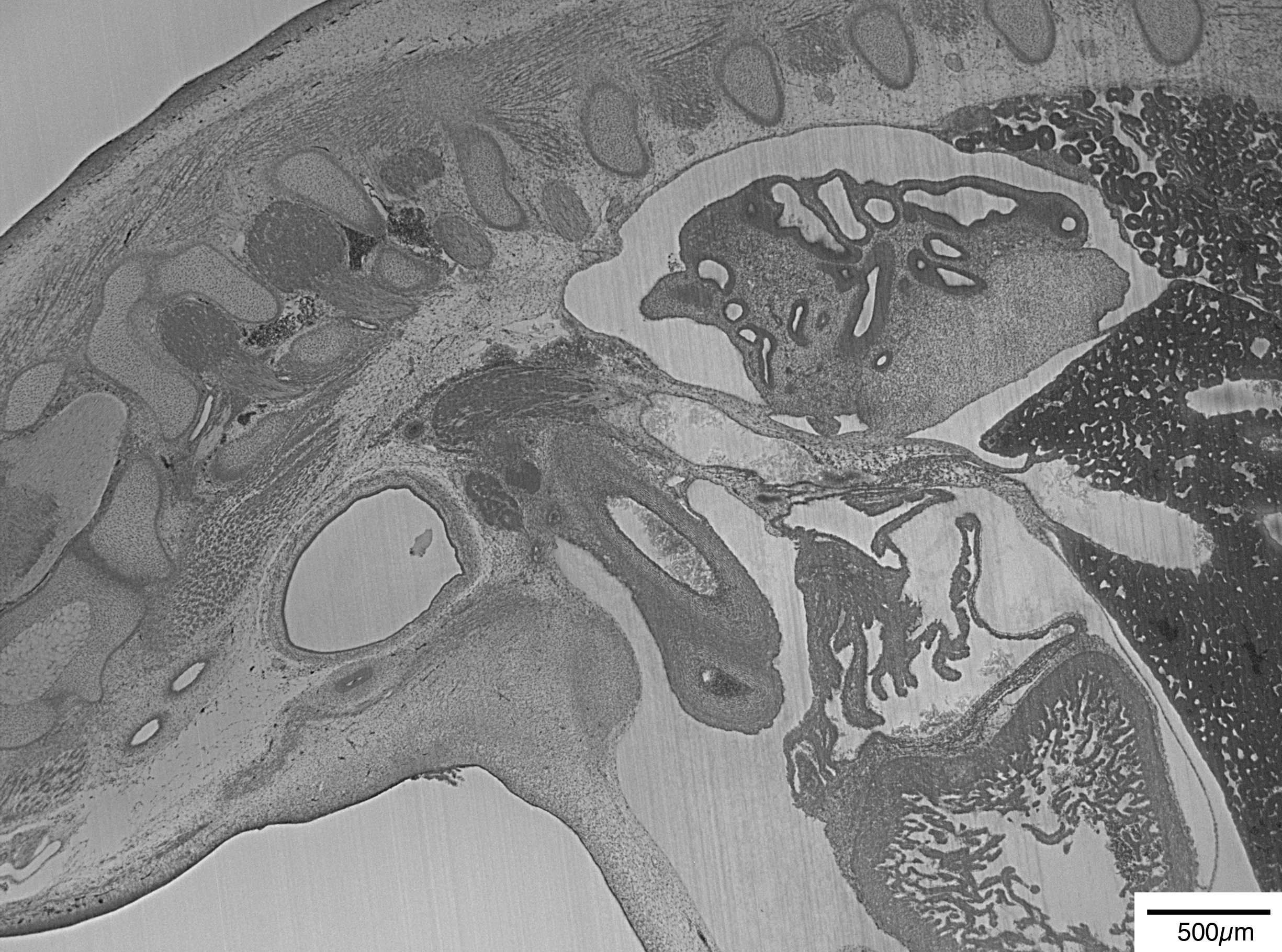
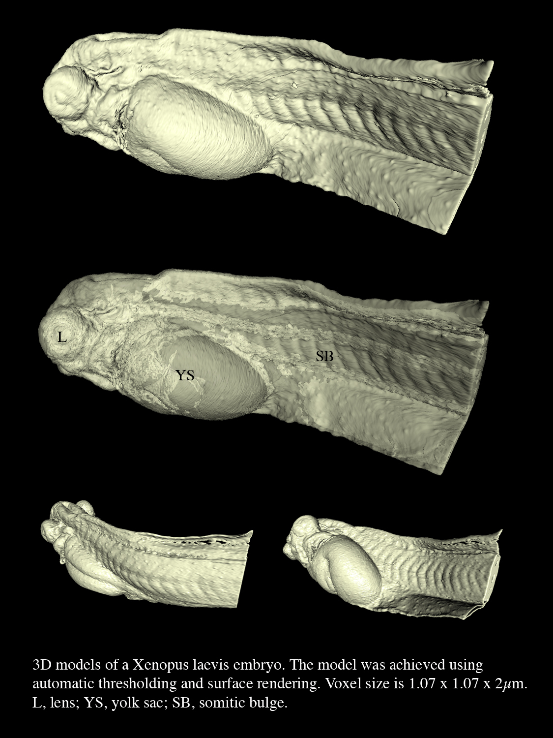
| Frog
(Xenopus laevis) Tadpole of approximately stage 36 |
Movie Grey-scale (jpg file) Colour jpg of 3D model |