
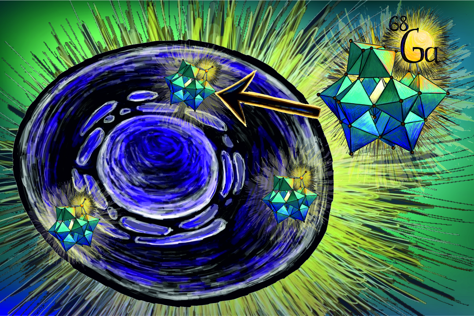
(Vienna, 17 November 2026) Understanding where cells used for therapeutic purposes distribute themselves within the body and how long they remain at their target site is of great importance for the development and optimisation of cell therapies. However, localising these cells in the body using imaging techniques remains a challenge. Researchers at the Medical University of Vienna and the University of Vienna have now developed a new radioactive marker that could, in future, help to visualise such cells using positron emission tomography (PET). The study results were published in the journal "Angewandte Chemie International Edition".
The new tracer, designated ⁶⁸GaPW₁₁, was developed as part of the "Joint Applied Medicinal Radiochemistry Facility", a collaboration between MedUni Vienna and the University of Vienna aimed at developing modern radioactive compounds for medical imaging. ⁶⁸GaPW₁₁ is based on a so-called Keggin polyoxometalate, an inorganic cluster of metal and oxygen atoms. The radioactive isotope gallium-68 is directly incorporated into the chemical structure. Unlike other tracers, the new tracer does not rely on binding to a specific receptor on the cell surface. As studies on a mammalian cell line show, it instead accumulates on the cell membrane and subsequently enters the cell interior. Furthermore, in cell experiments, GaPW₁₁ was taken up more readily by the mammalian cells under investigation than the reference compounds – a potential advantage for future cell imaging using PET.
"Polyoxometalates are fascinating inorganic compounds that we have been investigating for years to determine their biological activity. The fact that we can now label one of them directly with a clinically relevant radionuclide and track it within cells represents an important milestone for us – and opens the door to a completely new class of entirely inorganic radiotracers," says study leader Annette Rompel (Institute of Biophysical Chemistry, University of Vienna). "What particularly fascinates us is the fact that ⁶⁸GaPW₁₁ can cross the cell membrane and accumulate intracellularly," adds co-author Nadiia Gumerova, also from the Institute of Biophysical Chemistry at the University of Vienna.
It is this direct labelling of cells that could, in future, enable the tracking of the distribution of cells used for therapeutic purposes using PET. From a radiochemical perspective, it is crucial that the tracer can be produced reliably and with a high degree of purity. "Both of these criteria were met by ⁶⁸GaPW₁₁. At the same time, the structure proved to be stable during radiolabelling," says co-author Karsten Bamminger from the Department of Biomedical Imaging and Image-guided Therapy at MedUni Vienna, highlighting further advantages of the new tracer. "Here, we are combining inorganic chemistry with radiochemistry and cell biology. This opens up the possibility of specifically utilising the properties of the chemical structure for cell imaging," summarises Markus Mitterhauser (Department of Biomedical Imaging and Image-guided Therapy, MedUni Vienna), coordinator of the research collaboration.
Cell therapies are becoming increasingly important in medicine. In particular, treatments are already being used for certain types of cancer in which immune cells are deployed specifically against tumour cells. Further cell therapies are being researched for various diseases. Methods that allow the distribution of therapeutically administered cells within the body to be tracked are of great importance for the development and optimisation of cell therapies. The new gallium-68 tracer is, as yet, a preclinical research approach; its suitability for PET imaging in living organisms is to be investigated in further studies.
Publication: Angewandte Chemie International Edition
A ⁶⁸Ga-Substituted Keggin Phosphotungstate as an Intrinsically Radiolabelled Inorganic PET Tracer for Cell Tracking.
Ella Duvanova, Joshua Rieger, Karsten Bamminger, Nadiia I. Gumerova*, Markus Mitterhauser, Annette Rompel*.
https://doi.org/10.1002/anie.9529520
This work was funded by the Austrian Science Fund (FWF) and would not have been possible without this funding, as well as the resources and collaboration of the University of Vienna and the Medical University of Vienna.