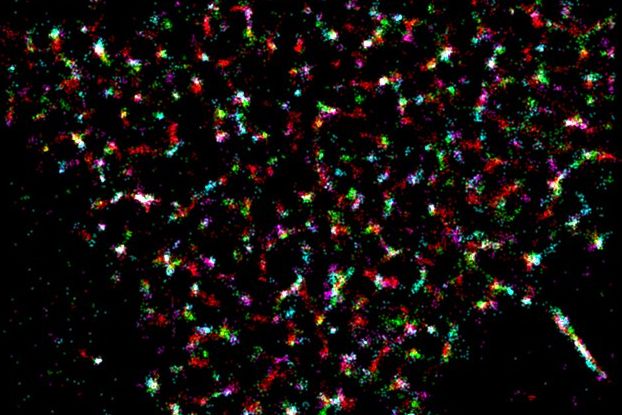
(Vienna, 14 June 2016) What were previously thought to be nanoprotein clusters on cell surfaces are often merely multiple counts. A method developed by Vienna University of Technology in collaboration with MedUni Vienna is now able to exclude artefacts and to examine cells nanoscopically.

Using light, it is not possible to image structures smaller than half the wavelength – at least that was what we believed for a long time. However, the development of so-called nanoscopy has shown that this rule has certain loopholes. If different molecules are lit up at different times, they can ultimately be joined together to produce a sharp image. The Nobel prize for chemistry was awarded for this in 2014. Since then, nanoscopy has become a globally applied method, which is used to examine the structure of the cell membrane, amongst other things. However, these methods are very laborious and prone to errors and often lead to false conclusions. A new method that has recently been published in the leading journal "Nature Methods" by a team from Vienna University of Technology and MedUni Vienna, is able to filter out artefacts and recognize real nanostructures.
Nano cell imaging techniques for medical applications
"For many biological or medical problems, it is crucial to understand the exact structure of the cell membrane," says Florian Baumgart from the biophysics research group led by Gerhard Schütz at the Institute for Applied Physics at Vienna University of Technology. Nanoscopy is an ideal tool for examining the spatial arrangement of proteins on the cell membrane. This first opportunity to explore the nanoarchitecture of cells will provide new insights into the way cells function. "We are particularly interested in the nanoarchitecture of T-cells and antigen-presenting cells in the initiation of the adaptive immune response," says project partner Hannes Stockinger from the Institute for Hygiene and Applied Immunology at MedUni Vienna's Center for Pathophysiology, Infectiology and Immunology.
The researchers are confident that they will be able to refine these novel methods, which allow cell resolution of less than 50 nm and even living cell analysis within microseconds, for the purposes of precision diagnostics. By mapping individual nanostructures and reactions, they hope to be able to create a new personalized biomarker profile, so that drug regimes can be specifically tailored to the individual in question.
Service: Nature Methods
Varying label density allows artifact-free analysis of membrane-protein nanoclusters
Florian Baumgart, Andreas M Arnold, Konrad Leskovar, Kaj Staszek, Martin Fölser, Julian Weghuber, Hannes Stockinger & Gerhard J Schütz – Nature Methods (2016).
http://dx.doi.org/10.1038/nmeth.3897