
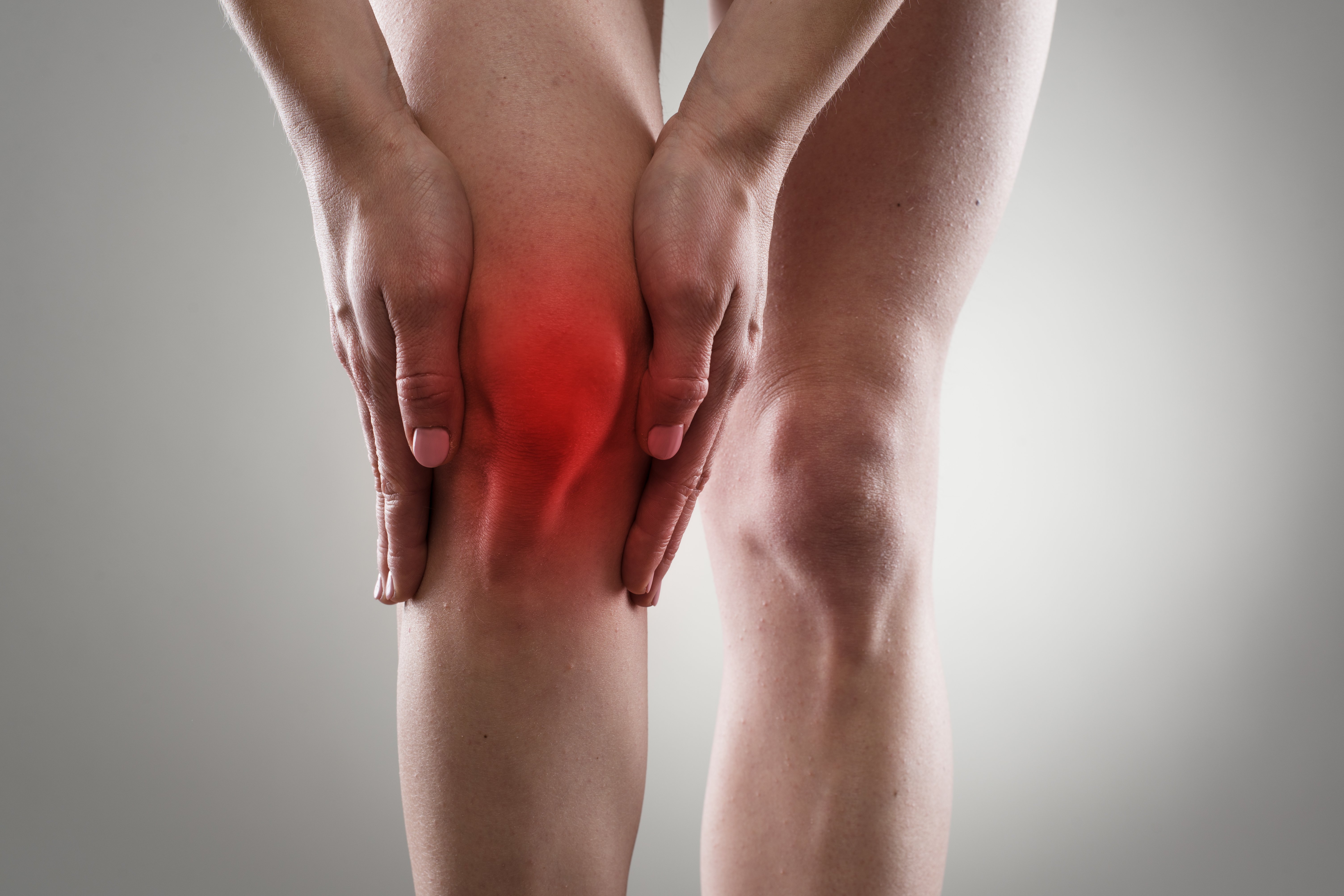
(Vienna, 08 October 2018) Patients frequently experience severe chronic pain following knee operations. Although the pain is thought to be due to damage to small nerves, it was hitherto impossible to demonstrate this by imaging. Two studies conducted by an interdisciplinary team of researchers led by radiologist Georg Riegler from MedUni Vienna’s Department of Biomedical Imaging and Image-guided Therapy have now successfully demonstrated these tiny, sensitive, cutaneous nerves on the anterior and medial thigh and around the knee using high-resolution ultrasound imaging. This means that targeted treatment can now be given. The studies have recently been published in leading journals "Arthroscopy" and "Ultraschall in der Medizin" [Ultrasound in Medicine].
Following a knee operation, patients often suffer months of chronic (and unexplained) pain, even though the operation itself was successful. In most patients, this pain is bearable and eventually disappears spontaneously. However, between 5 and 10% of patients are still suffering twelve to eighteen months later, in some cases with severe, so-called neuropathic pain. This naturally has a negative impact upon their quality of life. It is only when all other objective causes have been ruled out that nerve damage is suspected as the cause.
During surgical procedures it is inevitable that there will be damage to tissue and nerves at the site of the operation. However, in most cases this does not have any further consequences and the normal pain due to the operation subsides again after a few weeks. However, if the nerve damage is so severe that the pain persists months after the operation or even worsens, targeted pain therapy should be started as quickly as possible to prevent chronification of the pain. Should this happen, then it is usually much more difficult for patients to ever become completely pain-free.
However, it was previously impossible to demonstrate the tiny cutaneous nerves, which are less than 1-mm thick, around the knee, and their branches, by means of imaging techniques. Hence, nerve damage could only ever be presumed and treated based on a suspicion. However, should the assumption prove false, such treatment can do more harm than good. An interdisciplinary team led by radiologist Georg Riegler from MedUni Vienna's Department of Biomedical Imaging and Image-guided Therapy, in collaboration with PUC - Private Ultrasound Center – and the Division of Plastic and Reconstructive Surgery at MedUni Vienna's Center for Anatomy and Cell Biology has now managed to demonstrate these small cutaneous nerves on the knee for the first time using high-resolution ultrasound imaging. In the first study, the team managed to demonstrate the entire course of ramus infrapatellaris (a sensitive nerve branch in front of and below the kneecap) by means of high-resolution ultrasound imaging. The second study demonstrated the branched anterior cutaneous branches of the femoral nerve, nervus femoralis. It was also found that the paths of these nerves are highly variable and follow a different course in each person. It is therefore essential to determine the exact position of the damaged nerve fibres before giving any targeted treatment to combat nerve pain.
According to Riegler, to accurately isolate the nerve branches causing the pain, it is essential to carry out a so-called "diagnostic blockade": "Since the nerve supply is so variable, it is essential to first make sure which of these tiny nerves is causing the pain. This can only be done by selectively temporarily anaesthetising, or blockading, the suspected nerve, with a maximum of 1 ml of anaesthetic. If the pain is significantly reduced immediately after a blockade has been done and returns once the anaesthetic has worn off, then we have located the problem."
If a nerve is causing the pain, there are various treatment options available. The first option is to treat locally with pain patches or physiotherapy. The next stage would be ultrasound-guided treatment, in which the affected nerve is repeatedly blockaded with anaesthetic in several sessions, thereby blocking the pain. Other possibilities are cortisone injections or radiofrequency ablation. Surgical procedures to release or sever the nerve are only considered as a last resort. However, this is often very successful.
Says Riegler: "It is very important to have an accurate diagnosis as quickly as possible, without waiting too long. Otherwise, the pain can burn itself into the "pain memory" and become chronic." Therefore, in future, better help will be available to patients with nerve damage following knee operations.
Service:
Pivec1, Gerd Bodner1, Johannes A. Mayer2 , C. Brugger3, Istvan Paraszti3, VeithMoser4, Hannes Traxler3, Georg Riegler1: Novel Demonstration of the Anterior Femoral Cutaneous Nerves using Ultrasound
DOI https://doi.org/10.1055/s-0043-121628
Georg Riegler, M.D., Suren Jengojan, M.D., Johannes A. Mayer, M.D., Christopher Pivec, M.D., Hannes Platzgummer M.D., Peter C. Brugger, M.D., Ph.D., Oskar Aszmann, M.D., and Gerd Bodner, M.D.: Ultrasound Anatomic Demonstration of the Infrapatellar Nerve Branches.
DOI: https://doi.org/10.1016/j.arthro.2018.05.043