
(Vienna, 03 April 2019) A MedUni Vienna study group led by Peter Pietschmann from the Institute of Pathophysiology and Allergy Research in cooperation with the University of Vienna and the Museum of Natural History Vienna has made a detailed analysis of the microarchitecture of bones affected by the bone metabolic disease known as Paget’s Disease. High-resolution micro-computed tomography techniques offer insights into the course of the disease.
Up to 5 – 8% of people in their 80s can be affected by the bone metabolic disease osteodystrophia deformans (Paget’s Disease). As a rule, Paget’s Disease occurs after the age of 55 and, as a result of disorganised bone remodelling, gradually causes sclerosis (thickening) of the bone tissue, which is often accompanied by bone deformation.
Although Paget's disease of the bone (PDB) is the second most common metabolic bone disease, there is limited information available about the microarchitecture of the affected bones, one of the main determinants of bone strength. Indeed, the microstructure of long, weight-bearing bones has never been systematically analysed.
The aim of the study was therefore to determine the cortical and trabecular bone characteristics at clinically relevant sites using micro-computed tomography (μCT).
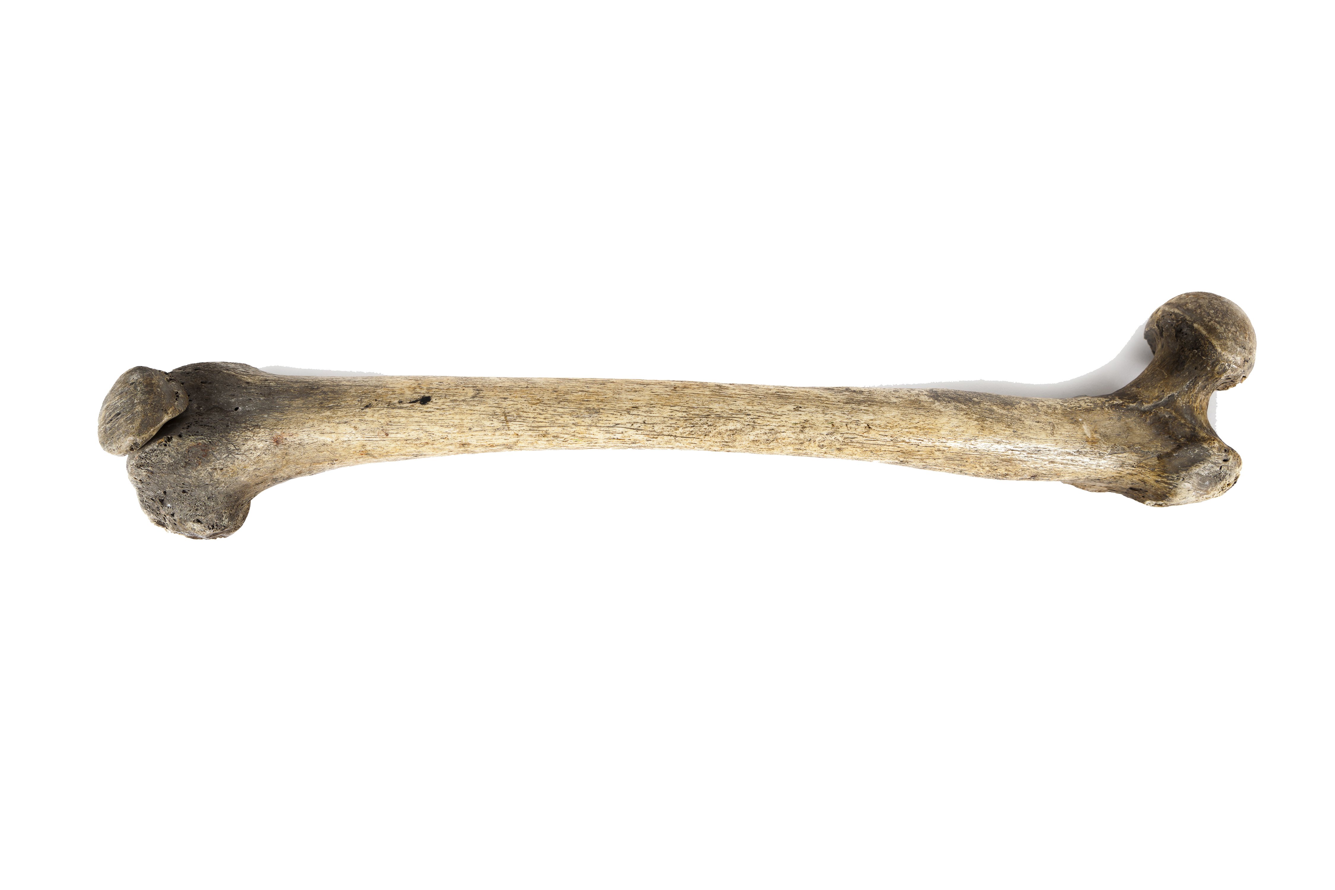
As part of a collaboration between the Medical University of Vienna, the University of Vienna and the Museum of Natural History Vienna, μCT analyses were carried out on historical femora (femurs) and tibiae (tibias). In the study, a comparison was made between 10 femurs and 10 tibias from the Museum of Natural History Vienna, which had been affected by the disease, and 13 femurs and 10 tibias from unaffected donors. The cortical and trabecular bone microarchitecture was digitised using an X-ray based μCT scanner.
Higher risk of bone fractures despite thickening
Increased cortical thickness, cortical porosity and trabeculation of the cortical structures were observed in the cortical compartments. Serious defects, disruption of the trabecular structures and increased thickness of the trabeculae were observed in the trabecular compartments.
These findings are relevant for differential diagnosis and bone fragility in Paget’s Disease, explains principal investigator Peter Pietschmann: "Although cortical thickness is increased in Paget’s Disease, the disease carries an increased risk of bone fracture. By identifying increased cortical porosity, our study has provided an explanation for increased bone fragility. In addition to this, our data are also significant for the diagnosis of Paget’s Disease in both historical and contemporary bone samples."
Service: Calcified Tissue International
Paget’s Disease of Long Bones: Microstructural Analyses of Historical Bone Samples
Elena Nebot, Patrick Heimel, Stefan Tangl, Martin Dockner, Janina Patsch, Gerhard W. Weber, Michael Pretterklieber, Maria Teschler Nicola, Peter Pietschmann; Calcified Tissue International 2019
https://doi.org/10.1007/s00223-019-00539-8