
(Vienna, Cambridge, 31 January 2020) A new imaging technique makes it possible to observe, in real time, which regions of a tumour are active. In combination with genetic tests, this could allow better personalised treatments and earlier assessment of their efficacy. The first study, which used this technique to investigate different types of breast cancer, has been carried out at the University of Cambridge by researchers led by Ramona Woitek from MedUni Vienna's Department of Biomedical Imaging and Image-guided Therapy and Ferdia Gallagher (Adjunct Professor at the Meduni Vienna)and was recently published in the specialist journal "PNAS".
The new imaging technique is based on Magnetic Resonance Imaging (MRI). This provides high-resolution images of organs and even tumours but has the disadvantage of not being very sensitive. For years now, scientists have been looking for ways to increase the sensitivity of the technique. For example, substances that only occur in low concentrations in the body are made visible so that metabolic processes can be tracked in real time.
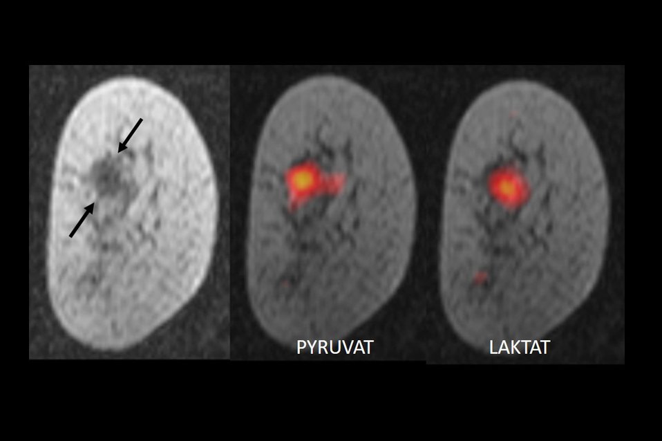
The group at the University of Cambridge labelled pyruvate, a substance that occurs naturally in the body, with the non-radioactive carbon isotope C-13 and then magnetised it. To do this the labelled pyruvate is cooled to -272°C and exposed to very strong magnetic fields and to microwave radiation – this process is also known as "hyperpolarisation". Once the substance has been thawed and dissolved, it is injected into patients. Due to the magnetisation, the signal strength of the C-13 pyruvate in MRI is increased 10,000-fold so that MR images of the tumour metabolism can be produced within seconds. Normally, the body's cells convert pyruvate into lactate. Due to their altered metabolism, tumour cells convert pyruvate to lactate much more rapidly – the speed of the process varying between different tumours and also within the tumour itself. Using MRI, it is not only possible to observe the quantity of the specially treated pyruvate in different tissues but also to observe the conversion process in real time. The researchers demonstrated that this provides information about the nature of the tumour and how aggressive it is.
"This is one of the most detailed images of the metabolism of breast cancer that we have ever had. It is as if we can see the tumour breathing," explains Kevin Brindle from the Cancer Research UK Cambridge Institute, one of the originators of the technique, in a press release.
So far the researchers have used the technique on seven patients with different breast cancers and it has enabled them to determine the nature and aggressiveness of the tumour. The technique is now going to be trialled on a larger group and also used to determine the treatment response of individual patients at an earlier stage by observing metabolic changes, thus ensuring that patients can receive the ideal treatment for them as early as possible.
Service: PNAS
Imaging breast cancer using hyperpolarized carbon-13 MRI
Ferdia A. Gallagher, Ramona Woitek, Mary A. McLean, Andrew B. Gill, Raquel Manzano Garcia, Elena Provenzano, Frank Riemer, Joshua Kaggie, Anita Chhabra, Stephan Ursprung, James T. Grist, Charlie J. Daniels, Fulvio Zaccagna, Marie-Christine Laurent, Matthew Locke, Sarah Hilborne, Amy Frary, Turid Torheim, Chris Boursnell, Amy Schiller, Ilse Patterson, Rhys Slough, Bruno Carmo, Justine Kane, Heather Biggs, Emma Harrison, View ORCID ProfileSurrin S. Deen, Andrew Patterson, Titus Lanz, Zoya Kingsbury, Mark Ross, Bristi Basu, Richard Baird, David J. Lomas, Evis Sala, James Wason, Oscar M. Rueda, Suet-Feung Chin, Ian B. Wilkinson, Martin J. Graves, Jean E. Abraham, Fiona J. Gilbert, Carlos Caldas, and Kevin M. Brindle
PNAS first published January 21, 2020; https://doi.org/10.1073/pnas.1913841117