
Percutaneous pulmonary valve replacement / Melody valve
The implantation of a heart valve using catheter technology is one of the latest advances in interventional cardiology. At present, the technique is mainly used to replace the pulmonary valve (pulmonary artery valve), while the aortic valve is mainly replaced in adults.
The following explanations apply to the much more frequently used "Melody valve", the schematic illustrations are taken from the Medtronic brochure/website.
What is a percutaneous pulmonary valve?
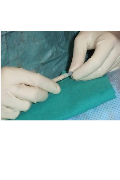
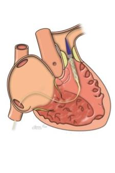
These are heart valves that are prepared in such a way that they can be sewn into a stent . This stent with the valve inside can then be attached to a balloon catheter. During implantation, the balloon is filled and presses the stent with the valve against the outlet part of the right ventricle - where the patient's own heart valve (pulmonary valve) no longer functions.
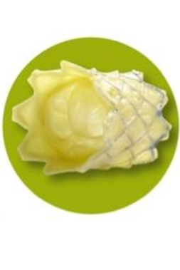
The heart valve itself comes from the calf's jugular vein, which is very well suited as a pulmonary valve - incidentally, the same valve is used by surgeons in the operating theatre when a valve replacement has to be performed on young children.
The stent is made of the metal platinum-iridium, which has very good erection forces and is very well contrasted in fluoroscopy. The stent and thus the valve can be widened to a diameter of 22 millimetres, which corresponds to the normal size of a pulmonary valve in adults. There are no known rejection reactions against the valve or the stent.
In 2000, paediatric cardiologist Philipp Bonhoeffer implanted a heart valve in a child for the first time during a cardiac catheter examination. The procedure is now accepted as a therapy and represents a good alternative to surgery.
The most widely used implant is the „Melody valve“ from Medtronic, as it can be used in a wide age range from children to adults. The Sapien valve from Edwards is also used in adults; it has a slightly different design, but is basically also a stent valve that is implanted using a similar technique.
Who can get a "catheter valve?
The current recommendations for a percutaneous pulmonary valve are
- Patients following surgery in which their own pulmonary valve has been replaced by a homograft, a Contegra valve, a xenograft or a valve-bearing conduit.
Patients with pulmonary stenosis or pulmonary insufficiency or a combination of both are eligible. As the materials, in particular the introducer sheaths, are quite thick, children weighing less than 30 kg are only suitable for this intervention in exceptional cases.
Basically, the procedure is the same as any other cardiac catheterisation. The implantation of a pulmonary valve can also be performed during deep sleep.
The individual steps of implantation
- Determination of the pressure values in the right heart by direct measurement in the right ventricle and the pulmonary artery
- Angiography (contrast imaging) of the right ventricle and the pulmonary artery - to estimate the size ratios
- Angiography (contrast imaging of the coronary arteries) - to make sure that the pulmonary valve is not compressing the coronary arteries and that there is no circulatory disturbance of the heart.
- Balloon dilatation / balloon sizing of the old pulmonary valve - this allows pre-existing stenoses to be widened and the diameter of the future new pulmonary valve to be determined.
- Pre-stenting - To ensure that the catheter valve can be anchored well later and is not damaged in the usually calcified structures of the heart, in recent years the region in which the future valve is to sit has been reinforced in advance with metal vascular supports (stent). Usually only 1 stent is necessary, but several may be required.
- Implantation of the valve: The pulmonary valve is then implanted in such a way that it lies exactly in the previously inserted stents. The implantation catheter is then removed.
- Final angiography: The examination ends with the injection of a contrast medium into the pulmonary artery to assess the proper functioning of the new valve.
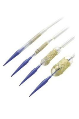
Mechanism of deployment of the Melody valve From top to bottom in the picture, the Melody valve is shown in the implantation system as it is pushed into the heart through the inguinal vein - then a protective sleeve is removed - and then a smaller balloon is filled, which expands the stent valve by about half -- finally, the stent is completely deployed by filling the large balloon.
How long does the flap work?
So far, the experience goes back twelve years. There are patients for whom the Melody valve has been working for over 10 years. In some patients, however, a second valve has had to be inserted into the first. This is due to weak points in the area of the stent (stent fractures). Although these have been reduced by pre-stenting, they still occur occasionally.
If a second valve should not or cannot be implanted, the catheter valve must be removed and a new valve surgically inserted.