Catheter-interventional closure of an arterial duct is now the international standard and can be performed at low risk from infancy. The newer implants even allow treatment of premature babies from approx. 2 kg body weight.
The intervention begins with visualisation of the duct using an injection of contrast medium. A distinction is made between different types of duct.
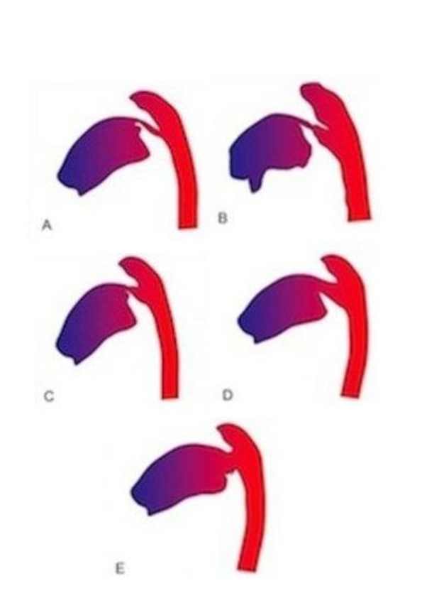
Different duct morphologies that influence the choice of implant:

A Small tubular duct without ampulla (implant cannot be placed completely in the duct)
B Tubular duct with ampulla (well suited for implantation of coils)
C Conical duct with ductal ampulla and pulmonary constriction (also well suited for coil closures but also for occluders)
D Large conical duct (usually multiple coils or an occluder required),
E "Window-like" duct (unsuitable for coils due to high risk of embolisation)
(Source: Pädiatrische Praxis, 2007, MICHEL-BEHNKE, I. The interventional closure of the ductus arteriosus. Indication, technique and innovations of a standard procedure)
After visualisation, the duct is measured precisely and the appropriate implant is selected. Various closure systems such as coils or occluders are available for this purpose.
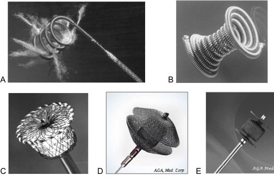
Coil closure systems

A Cook spiral (coil) reinforced with Dacron threads,
B Duct occluder coil (PFM),
C Amplatzer duct occluder (ADO I), a nitinol mesh with retention disc and plunger for transvenous closure of large ducts
D Amplatzer duct occluder (ADO II) with 2 retention discs,
E Amplatzer Duc
(Source: Pädiatrische Praxis, 2007, MICHEL-BEHNKE, I. The interventional closure of the ductus arteriosus. Indication, technique and innovations of a standard procedure)
Depending on the implant selected, it is inserted from the aorta or the pulmonary artery. The implants are attached to a retaining cable during placement and their position can be corrected if necessary. Only at the very end is the retaining mechanism unscrewed and the implant remains in place. Finally, a new injection of contrast medium is used to confirm the correct fit.
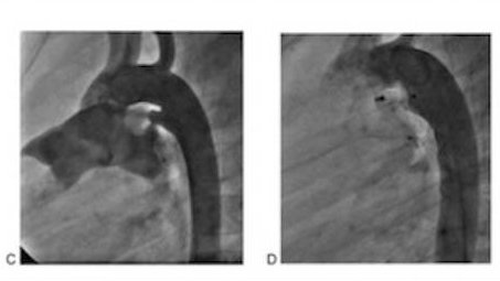
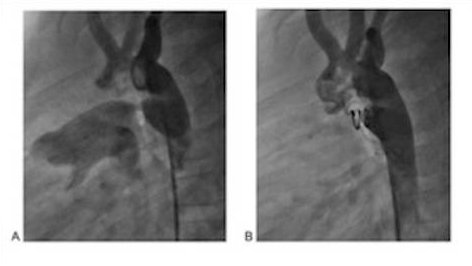
Duct closure using coil and occluder, angiographies in the lateral beam path:
A Tubular duct without large ductal ampulla
B Complete closure by implantation of a coil.
C Large conical duct ,
D Complete closure after implantation of an Amplatzer duct occluder.
(Source: Pädiatrische Praxis, 2007, MICHEL-BEHNKE, I. The interventional closure of the ductus arteriosus. Indication, technique and innovations of a standard procedure)

