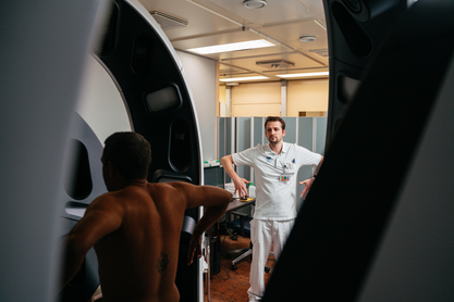
(Vienna, 27 June 2024) The Department of Dermatology at University Hospital Vienna and MedUni Vienna is using a whole-body imaging system. Compared to the previously used 2D full-body photography, which takes up to 20 minutes, it is now possible to capture almost the entire skin surface in macro resolution within a few seconds. The system supports doctors in differentiating melanomas from other skin lesions and enables the diagnosis of melanomas at very early stages.
In addition to the ultra-high-resolution 3D overview image of the entire body, the integrated digital handheld device (=digital dermoscopy) can be used to take individual detailed images at 15-200x magnification. Overall, however, only a few such individual images are required with the device, which also increases comfort for the patient.
Skin changes can be marked using the integrated software and observed over time. All overview and close-up images as well as searchable features and notes are managed in a secure image management system.
In addition to the examination of melanomas, the system can also be used for the documentation of inflammatory and pigmented skin lesions. These include psoriasis and vitiligo, for example, but the system can also be used for the treatment of lymphoedema or burns.
AI solution being tested
An innovative AI-controlled evaluation system for suspicious lesions is also integrated into the device. The artificial intelligence was previously trained using more than 66,000 images annotated by doctors. The newly recorded lesions of patients are grouped, measured and compared in order to detect developing or changing lesions at an early stage.
A clinical study has already shown that the automated analysis of whole-body 3D images can distinguish melanoma from other skin lesions with a high degree of accuracy.