
(Vienna, 10 May 2017) The Department of Pathology at the Medical University of Vienna is a partner in the recently launched EU ChipScope research project to develop a new, extremely small and ultra-high-resolution light microscope. This should enable us to observe the inside of living cells in real time. Seven institutes in five European countries will be taking part in this extremely technically demanding project over the next four years.
The potential future applications of the newly developed microscope, which is only a few millimetres in size, are extremely diverse. The project partners are each focusing on a specific application. The project will use medical laboratory tests with the new microscope to observe changes inside living cells in real time. The experiments will focus on examining cells of patients with idiopathic pulmonary fibrosis (IPF). IPF is an aggressive and rapidly progressing lung disease that kills half a million people each year worldwide.
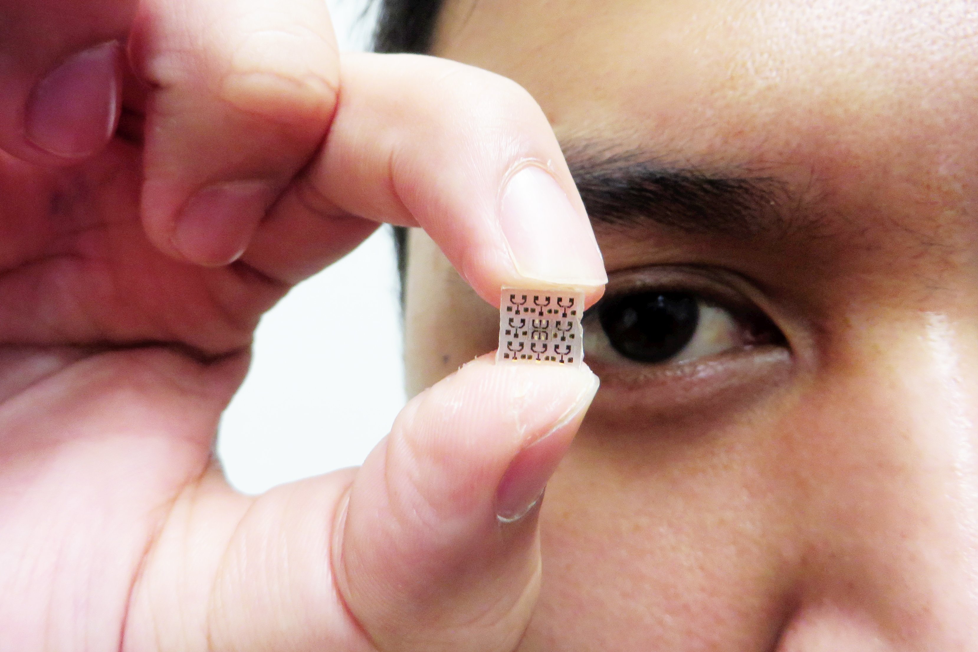
The new microscopes will be inexpensive and easy to use. This will facilitate and speed up research results in the fields of medicine, biology and biotechnology, as well as many other areas. The compact microscope will also be very attractive to developing countries, since they will be able to make cheap high-resolution microscopes locally in order to identify pathogens, for example. In the not too distant future ChipScope microscopes might also be incorporated in consumer electronics, in the same way as smartphone cameras are today.
Why is there a need for new optical microscopes?
What we can currently see with conventional optical microscopes is limited by the laws of physics, or more precisely the wavelength of light, which is approximately half a thousandth of a millimetre. However, cell components, DNA molecules and proteins are much smaller and cannot therefore be seen with these conventional microscopes. The aim is therefore to develop very small LEDs with a diameter of only 50 nm (that is 1000x smaller than the diameter of a human hair) and use them as light sources for a new microscope. What distinguishes them from conventional optical microscopes is that the illumination comes from extremely small, individually regulated light sources and not from a wide illumination area and tiny detectors in the camera. This allows the reconstruction of a transmitted light image and hence real-time high-resolution optical microscopy of extremely small structures such as bacteria or processes inside living cells.
The project team led by Silvana Geleff at the Department of Pathology will produce cell cultures from the lungs of patients with idiopathic pulmonary fibrosis and from the lungs of healthy donors and test the optimum growth conditions for the experiments with the new microscope. Geleff and her colleagues Sigurd Krieger and Nicole Huttary will be working closely with Viennese project partner AIT to optimise the use of the ChipScope to detect intracellular processes under physiological and pathological conditions and to establish this use for subsequent routine application.
Highly specialised, interdisciplinary project team
The ChipScope project runs from January 2017 until December 2020. The project partners are SMEs, universities and research institutes from five European countries: Braunschweig University of Technology, Tor Vergata University in Rome, the company Expert Ymaging in Barcelona, the Austrian Institute of Technology AIT in Vienna, the Department of Pathology of the Medical University of Vienna and the Swiss Foundation for Research in Microtechnology FSRM. The project is being coordinated by the University of Barcelona.