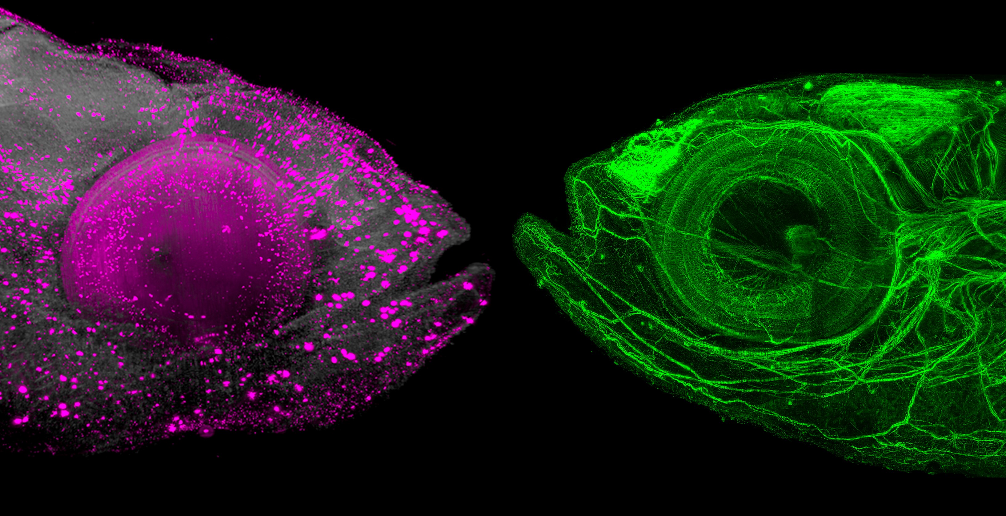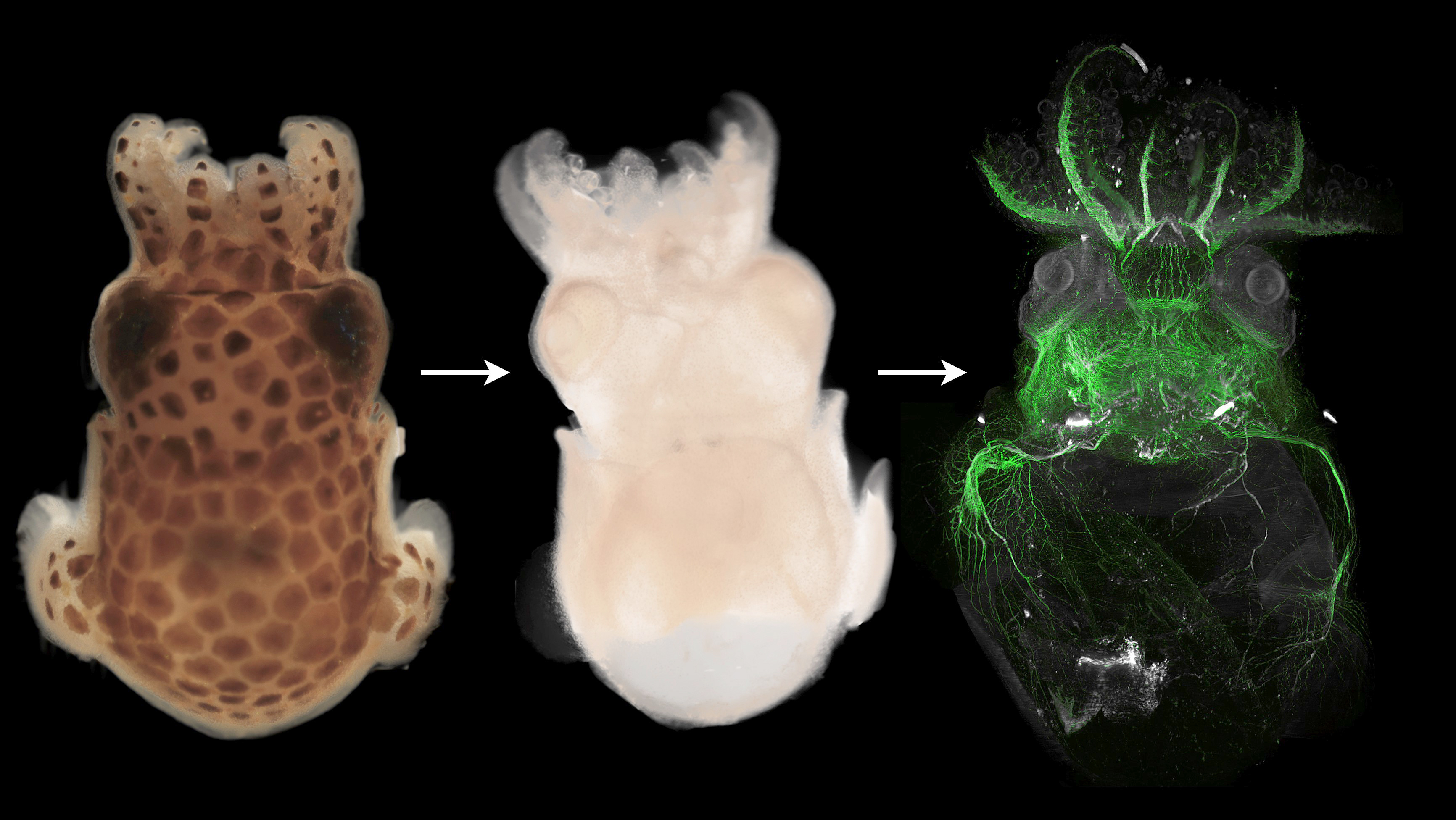
(Vienna, 02 June 2020) Spectacular images are possible using a new method developed by Vienna University of Technology and Max Perutz Labs, a joint venture of the University of Vienna and the Medical University of Vienna. Animals are made transparent so that you can see inside them.
What actually goes on inside a bristle worm? How are specific types of cells distributed inside the body of an axolotl? What does the neuronal structure of a zebrafish look like? Up until now, there was only one possible way of answering biological questions such as this: cutting animals up into slices and examining them under a microscope. However, many important structures are destroyed in the process.
A much more elegant option is so-called "clearing". This involves making the animals transparent using special chemical techniques. Certain types of cells are marked with special fluorescent dyes. The animal is then illuminated using an "ultramicroscope", producing spectacular images that carry a vast amount of valuable biological information.
There has now been an important breakthrough in this process. Whereas, previously, the clearing techniques that were used were only optimised for a specific type of tissue, e.g. mouse brains or fruit flies, an improved technique has now made it possible to render any animal completely transparent and to examine it microscopically.
The project was coordinated by research group leader Florian Raible (Max Perutz Labs) and involved MedUni Vienna's Center for Brain Research (CBR) and the Research Institute for Molecular Pathology (IMP), as well as Vienna University of Technology (TU Wien). The paper has now been published in "Science Advances".
Clearing the pigments
Vienna University of Technology has been researching ultramicroscopy for many years now. In this technique, biological tissue is illuminated and recorded layer by layer, using a thin sheet of laser light. From these images, a three-dimensional model of the animal is then generated on a computer. "In this way, it possible to overcome many optical limitations that one encounters in other techniques. The light sheet enables us to produce high-resolution images extremely quickly," says Saideh Saghafi, who designed the microscope at Vienna University of Technology.

However, in order to be able to do this, it is first of all necessary to modify the tissue so that the laser beam can penetrate it. "The first step is to clear the biological tissue – to make it transparent," explains the developer of the clearing technique, Marko Pende from the Institute of Solid-State Electronics of Vienna University of Technology and the CBR. One of the great challenges in doing this is that an organism contains a whole range of pigments, which first have to be broken down. "However, we have now discovered that this can be done really quickly with a cleverly chosen combination of chemicals and, indeed, for many different types of animal."
It is therefore now possible to examine different types of animal – from molluscs via bony fish right through to amphibians. "These are just a few examples. We are assuming that the technique will work for many different animals, but we have not yet tested it on all of them," explains Hans Ulrich Dodt, senior author of the publication.
Fluorescent markings
If one wants to look at highly specific structures or cells, these can be marked with fluorescent molecules. As soon as they are hit by the laser beam, they emit light and become visible. It is therefore possible to map three-dimensional structures inside the animal or to examine the spatial distribution of certain cells or molecules within the body.
The new technique allows these markings to be affixed to different biomolecules: "You can work with genetic techniques, introduce marked antibodies against specific proteins into the tissue or mark specific RNA molecules," says Florian Raible (Max Perutz Labs). "Due to the wide range of potential applications, our technique is of interest for many different fields of research."
However, it must be borne in mind that the marker molecules do not penetrate all types of tissue at the same rate. Hitherto, it was hardly possible to reach difficult-to-access points deep inside an animal. Also, in some cases, the chemicals used to destroy the pigments also damaged the fluorescent markers.
The recent research project managed to solve all these problems. "Our technique now enables us to illuminate entire cell networks in the animal and map them three-dimensionally," reports Marko Pende.
"This is a new and powerful examination technique for the field of biology," emphasises Florian Raible. "We are confident that it will enable us to answer important questions in biological research that have hitherto defied detailed investigation."Original publication"A versatile depigmentation, clearing and labeling method for exploring nervous system diversity", Science Advances, DOI: 10.1126/sciadv.aba0365