
Wir erforschen neue MR-Bildgebungsmethoden, um Stoffwechselveränderungen im Gehirn und in anderen Organen zu erkennen.
Unsere Arbeitsgruppe erforscht neue Methoden der Magnetresonanz-spektroskopischen Bildgebung, um Stoffwechselprozesse im menschlichen Körper nicht-invasiv, räumlich aufgelöst und ohne radioaktive Tracer zu untersuchen.
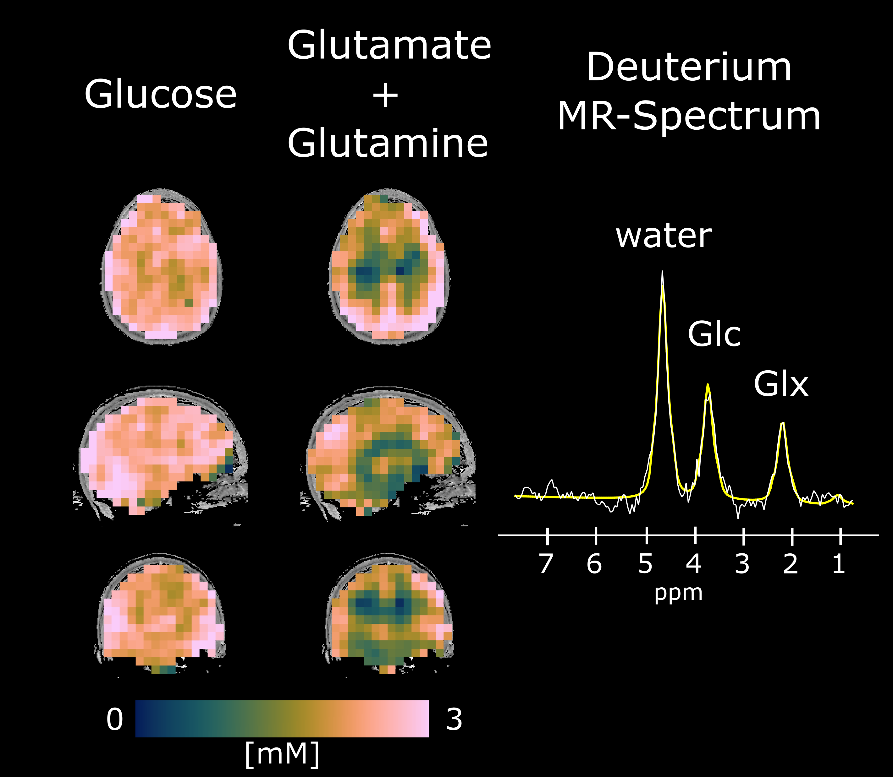
Ein Schwerpunkt liegt auf Deuterium Metabolic Imaging (DMI) und ¹H-MR-spektroskopischer Bildgebung (¹H-MRSI) nach Gabe deuterium-markierter Substrate. Damit lassen sich Glukoseaufnahme und nachgeschaltete Stoffwechselprodukte wie Glutamat/Glutamin dynamisch im Gehirn abbilden. Ziel ist es, metabolische Veränderungen bei neurologischen und onkologischen Erkrankungen besser zu verstehen und neue MR-Biomarker für Forschung und Klinik zu entwickeln.
Wir kombinieren schnelle 3D-MRSI-Akquisitionen, ultra-hochfeld MR bei 7 Tesla, nicht-kartesische k-Raum-Trajektorien, quantitative Modellierung und moderne Rekonstruktionsmethoden. Ein besonderer Fokus liegt auf der Translation dieser Methoden in klinisch relevante Anwendungen, etwa zur Untersuchung von Hirntumoren, neurodegenerativen Erkrankungen und systemischem Stoffwechsel.

Fabian Niess
GruppenleiterDipl.-Ing., PhD
ORCID: 0000-0003-1235-7595
T +43 1 40400-37720
fabian.niess@meduniwien.ac.at
- MRT des dynamischen Gliom-Stoffwechsels (2022)
Klinische Forschung FWF 399.998 € PI: Fabian Niess - Zusammenhang zwischen Gewichtsverlust und Gehirnstoffwechsel (2026)
Klinische Forschung FWF 445.890 € PI: Fabian Niess - GLUCOSCAN (2023): Deuterierung zur Verbesserung der Spezifität und Empfindlichkeit der Magnetresonanztomographie bei der Krebsdiagnostik
European Research Council (ERC) 2.495.924 € PI: Wolfgang Bogner
- Bader V et al. Assessment of T1 and T2 relaxation times of deuterium (2H) labeled resonances in the human liver and kidney using k-space reordered 3D concentric ring trajectory sampling at 7T (Magnetic Resonance Materials in Physics, Biology and Medicine, 2026)
- Niess F et al. Advanced methods in deuterium metabolic imaging (Magnetic Resonance Materials in Physics, Biology and Medicine, 2026)
- Niess F et al. Feasibility of High-Resolution Deuterium Metabolic Imaging of the Human Kidney Using Concentric Ring Trajectory Sampling at 7T (NMR in Biomedicine, 2025)
- Frese S et al. Balanced Steady-State Free Precession Enables High-Resolution Dynamic 3D Deuterium Metabolic Imaging of the Human Brain at 7T (Investigative Radiology, 2025)
- Bader V et al. Concentric Ring Trajectory Sampling With k-Space Reordering Enables Assessment of Tissue-Specific T1 and T2 Relaxation for 2H-Labeled Substrates in the Human Brain at 7 T (NMR in Biomedicine, 2025)
- Niess F et al. Whole-brain deuterium metabolic imaging via concentric ring trajectory readout enables assessment of regional variations in neuronal glucose metabolism (Human Brain Mapping, 2024)
- Niess F et al. Reproducibility of 3D MRSI for imaging human brain glucose metabolism using direct (2H) and indirect (1H) detection of deuterium labeled compounds at 7T and clinical 3T (NeuroImage, 2023)
- Niess F et al. Noninvasive 3-Dimensional 1H-Magnetic Resonance Spectroscopic Imaging of Human Brain Glucose and Neurotransmitter Metabolism Using Deuterium Labeling at 3T (Investigative Radiology, 2023)
- Bednarik P et al. 1H magnetic resonance spectroscopic imaging of deuterated glucose and of neurotransmitter metabolism at 7 T in the human brain (Nature Biomedical Engineering, 2023)