
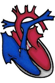
Aortenstenose (AS)
In this heart defect, the outlet from the heart into the main body artery (aorta) is narrowed. The most common form of aortic stenosis is narrowing of the aortic valve (valvular aortic stenosis). This narrowing can be caused by the leaflets of the aortic valve being too small or too thick or by a bicuspid aortic valve (two instead of three pockets), resulting in an impaired opening of the aortic valve.
Sometimes the narrowing does not affect the aortic valve itself, but there is a narrowing of the passage either above (supravalvular aortic stenosis) or below (subvalvular aortic stenosis) the aortic valve.
Welche Auswirkungen hat die Aortenstenose?
As the main pumping chamber, the left ventricle pumps oxygen-rich blood into the body through the aortic valve into the aorta. If the opening to the aorta is too narrow, the left ventricle has to work harder to pump enough blood into the body. This extra work causes the heart muscle to thicken (left hypertrophy).
Symptoms
Slight narrowing does not cause any symptoms. Even with higher-grade aortic stenosis, the patient is usually symptom-free, but the risk of serious cardiac arrhythmia is increased. This is particularly true during physical exertion, e.g. during sport, and in the worst case can lead to sudden cardiac death. In the case of subvalvular aortic stenosis, the aortic valve sometimes also develops a leak (aortic insufficiency).
How is aortic stenosis treated?
Valvular aortic stenosis: In the case of significant aortic stenosis, the opening of the valve leaflets is improved either surgically (commissurotomy - splitting of the valve suspension) or by cardiac catheterization (balloon dilatation) and the left main chamber is relieved. Which procedure is used depends, among other things, on whether aortic insufficiency is also present. Thanks to improvements in materials and special techniques, balloon dilatation using cardiac catheterization has replaced surgery in many centers. In the past, balloon dilatation was usually only performed on seriously ill newborns as an emergency procedure, but has now become established throughout childhood and adolescence. A catheter with a balloon is inserted into the heart and the balloon is inflated in the constricted valve to widen the narrowed opening of the valve leaflets. In the long term, most aortic valves become leaky (insufficient). Valve replacement surgery then becomes necessary, in which an artificial heart valve is inserted. Patients with an artificial valve receive lifelong anticoagulant medication (Marcoumar). In children, the so-called Ross operation is performed more frequently.
The aortic valve is replaced by the body's own pulmonary artery valve. In turn, the pulmonary artery valve is replaced by a donor valve. The donor valve may need to be replaced over time due to wear and tear or the patient's growth. For some patients, this replacement can already be carried out using cardiac catheterization (Melody valve).
Subvalvular aortic stenosis: In the case of a narrowing below the aortic valve, the surgeon removes the excess tissue. Cardiac catheter intervention is not possible with this form of aortic stenosis.
Supravalvular aortic stenosis: In the case of a narrowing above the aortic valve, the narrowed part of the aorta is surgically widened. Various techniques are used here, and it may also be necessary to implant a prosthesis (artificial vessel)
In the case of aortic stenosis, endocarditis prophylaxis is still recommended, especially after valve surgery.
Bei der Aortenstenose wird insbesondere nach Klappenoperationen eine Endokarditisprophylaxe weiterhin angeraten.