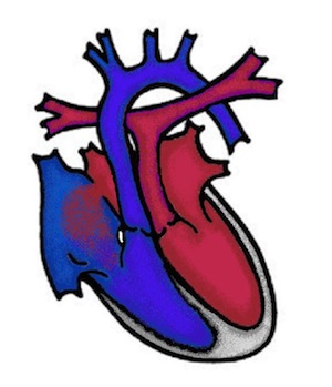
Transposition of the great arteries (TGA)
Die Transposition der großen Gefäße gehört zu den zyanotischen Herzfehlern (Zyanotische Herzfehler), bei dem die großen Gefäße (Hauptschlagader = Aorta und Lungenschlagader = Pulmonalarterie) vertauscht sind. Die Aorta entspringt aus der rechten Herzkammer, dadurch erhält der Körper nur das sauerstoffarme Blut. Die Pulmonalarterie entspringt aus der linken Herzkammer und bringt das bereits sauerstoffreiche Blut wiederum in die Lunge.
What are the effects of transposition of the great vessels?
Before birth, oxygen is supplied to the body via the placenta. After birth, the lungs develop and normally take over the oxygen supply. In TGA, however, there are two completely separate circulations: the oxygen-rich blood from the lungs cannot enter the systemic circulation and the oxygen-poor blood cannot enter the lungs. Survival is only possible if the short-circuit connections ductus arteriosus (PDA) and foramen ovale (PFO), which were present before birth, remain open.
Symptoms
The newborns stand out due to a blue coloration of the skin (cyanosis) and often have labored breathing. The first symptoms appear when the ductus arteriosus and the foramen ovale close, i.e. in the first three days of life.
How is transposition of the great vessels (TGA) treated?
Immediately after birth, the newborn is given medication (prostaglandin) to keep the ductus arteriosus (connection between the aorta and pulmonary artery) open and thus supply the lungs with deoxygenated blood.) If the foramen ovale (hole in the atrial septum) is too small, it is enlarged using a balloon (Rashkind procedure). These two connections allow the blood to be mixed and adequate circulation can be maintained.
Arterial switch surgery is usually performed in the first 5-10 days of life. This involves cutting off the pulmonary and aortic arteries above the valves and swapping them. The coronary vessels (coronaries) must also be replanted into the "new" aorta. This connects the aorta to the left main chamber and the pulmonary artery to the right main chamber.
In principle, the vast majority of patients are cured after an arterial switch operation. In the long term, constrictions can very rarely occur at the suture points and at the outlet of the coronary vessels. The constrictions at the sutures (supravalvular aortic or pulmonary stenosis) can usually be widened by balloon dilatation using a cardiac catheter intervention. As the coronary vessels are difficult to visualise with a cardiac ultrasound examination, a cardiac catheter examination is performed if a circulatory disorder is suspected. This allows the coronary vessels to be assessed in their entire course.
Additional anomalies
The form of TGA described here is the most common form of this complex cardiac malformation. However, there is also another variant, the corrected transposition of the great arteries (ccTGA). In this case, not only are the pulmonary and aortic arteries transposed to the corresponding ventricles, but also the atria to the ventricles. These double wrong connections cancel each other out, making this a form of TGA in which there is no cyanosis if there are no additional malformations. Both forms of TGA can also have ventricular septal defects, pulmonary stenosis or other anomalies and then require a different treatment than the one described here.
