
Pulmonary stenosis (PS)
Pulmonary stenosis is a narrowing of the pulmonary artery valve or the outlet from the right main chamber. There are 3 types of pulmonary stenosis:
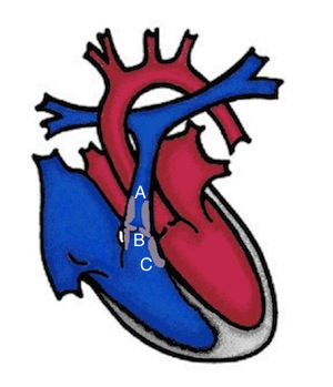
supravalvular PS - the narrowing is above the valve with a narrowing of the pulmonary artery (A);
valvular PS - here the valve itself is affected, the valve pockets are thickened or partially fused, the opening of the valve is not complete (B);
subvalvular PS (= infundibular PS) - the outlet from the right ventricle under the valve is narrowed by excess tissue (C).
What are the effects of pulmonary stenosis?
Pulmonary stenosis can be divided into low, medium and high severity depending on the degree of severity. The classification is based on the estimation of the pressure load on the right main chamber above the right ventricle. Medium to high-grade stenosis leads to enlargement and thickening of the right ventricle (right ventricular hypertrophy). The pressure in the right ventricle can become suprasystemic (higher than the pressure in the systemic circulation). The result is right heart failure.
How is pulmonary stenosis treated?
Minor pulmonary stenoses do not require treatment. The severity of pulmonary stenosis can increase or decrease with growth. Regular cardiac ultrasound checks are therefore necessary. If the pressure load in the right main chamber reaches a defined limit, treatment is necessary.
Supravalvular PS: The narrowed pulmonary artery can possibly be widened by balloon dilatation. In larger children or in the event of a renewed narrowing, the vessel can be kept open with a stent (wire mesh). If this is not possible or is unsuccessful, a surgical expansion with a patch is necessary.
Valvular PS: The valve is dilated in the cardiac catheterization laboratory using a balloon dilatation. This is the standard treatment worldwide for children and adults. Only in the case of severely malformed pulmonary valves is an operation necessary in which the opening of the stuck pocket valves is widened (commissurotomy). If this is not possible, a donor valve from a human (homograft) or from animal material (xenograft) is used. If there is also a leak in the pulmonary valve, a heart valve can also be implanted in older children using cardiac catheterization (Melody valve). In this procedure, a biological heart valve, which is sewn into a stent and mounted on a balloon catheter, is advanced into the heart via the inguinal vein and compressed at the site of the diseased pulmonary valve by expanding the balloon. This technique is also suitable for patients with tetralogy of Fallot or after Ross surgery if the surgical result deteriorates over the years.
Subvalvular PS: the excess tissue from the outflow tract of the right ventricle is surgically removed.