
We’d like to introduce you to the researchers behind the cancer research projects
100 percent of the donations raised by the Cancer Research Run go toward cancer research projects. Here, we’d like to introduce you to the researchers behind these projects and tell you what they’re currently working on.
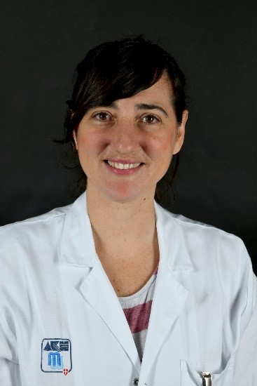
Verena Paulitschke of the University Clinic for Dermatology is investigating how blood proteins can predict the response to immunotherapies in metastatic melanoma. Her research could help personalize treatment, reduce side effects, and identify resistance mechanisms at an early stage.
What are you investigating in the study, and why is research in this area relevant?
Immune checkpoint inhibitors achieve excellent results in clinical practice; however, about half of patients exhibit intrinsic or acquired resistance. To date, there are no biomarkers that can accurately predict response to anti-PD-1 therapy in metastatic melanoma. Using mass spectrometry, we are analyzing the proteins in the blood of melanoma patients prior to immunotherapy and correlating these findings with treatment response to identify potential biomarkers. This approach could make it possible to stratify patients, avoid unnecessary side effects, save treatment time, and/or detect the onset of resistance at an early stage. In addition, our findings expand our understanding of the underlying resistance mechanisms, which could then be targeted with new treatment options.
What results have been achieved so far?
We have identified mechanisms that are upregulated in non-responders, including neutrophil degranulation, cell-substrate adhesion, and the organization of the extracellular matrix. Serum analysis led to a potential signature comprising 10 key markers. Analysis of primary melanoma cells from non-responders revealed a potential signature comprising 4 key markers. The data were published earlier this year in the renowned journal *Clinical Cancer Research* (PMID: 37861398) and also represent the core work of my PhD student, Nina Zila.
What does this mean for future research?
In the future, we plan to analyze additional patient samples not only before but also during therapy—on the one hand, to confirm and expand our existing signature of predictive markers, and on the other hand, to identify pharmacodynamic markers as well. In addition, we would like to use a completely novel method. To do this, we analyze patients’ finger sweat and, in collaboration with the Department of Analytical Chemistry at the University of Vienna (Christopher Gerner), determine the metabolites. This method is non-invasive, and sample collection is simple. This could enable the identification of biomarkers, as described in a recent study in *Nature Communications* (PMID: 34645808). Thus, it might be possible to identify metabolites that play a significant role in treatment response. If successfully established, the next step could be to implement this approach in clinical practice.
About the Author
Verena Paulitschke is a senior physician in dermatology at the University Clinic for Dermatology at the Medical University of Vienna. She studied medicine at MedUni Vienna and also completed her residency, PhD, and habilitation there. She completed a two-year research fellowship at ETH Zurich and the Department of Dermatology at the University Hospital of Zurich. She has been an associate professor since 2018. Her research focuses on evaluating resistance mechanisms to targeted therapies and immunotherapy, as well as identifying predictive markers in metastatic melanoma. She has already won numerous awards for her publications.
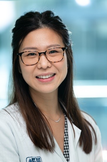
Are breast cancer patients with a BRCA1 gene mutation more likely to develop brain metastases? What other genes might be involved in this process, and would a new therapeutic approach be both feasible and successful? Yen Tan, a cancer epidemiologist at the University Clinic for Gynecology and a member of the Comprehensive Cancer Center (CCC) at MedUni Vienna and Vienna General Hospital (AKH), is investigating these questions in her project, which is supported by CCC research funding. In her study, she is conducting molecular genetic analyses of blood samples from patients to identify potential patterns of genetic changes. She hopes this will provide new insights into the signaling pathways involved in the development of brain metastases and, as a result, identify potential targets for new treatment methods.
Twenty to 30 percent of all patients with invasive breast cancer face a recurrence and the development of metastases. Five percent of these develop brain metastases, with the rate reaching up to 40 percent in certain subgroups of patients. This primarily affects women with HER2-positive or triple-negative breast cancer. Experts suspect that these numbers will rise, as patients are living longer due to improved therapies and will therefore reach the metastatic stage more frequently. New therapeutic approaches are thus urgently needed.
The majority of breast cancer patients with brain metastases are currently treated with local therapies (surgery and radiation oncology) and additional chemotherapy. Tan: “We want to investigate whether BRCA mutations occur more frequently in breast cancer patients who have developed brain metastases. If this proves to be the case, they could benefit from treatment with PARP inhibitors.”
PARP inhibitors are a class of drugs that block the repair of DNA damage in cancer cells, ultimately leading to their death. PARP inhibitors are usually used in combination with chemotherapy and are now considered the standard of care for cancers caused by BRCA mutations.
If other genetic mutations are involved in metastasis, new, personalized treatment approaches could also be developed in these cases.
About the Author
Yen Tan has been working at the University Clinic for Gynecology at the Medical University of Vienna since 2015 and is the deputy director of the Center for Hereditary Breast and Ovarian Cancer. She completed her postdoctoral training in molecular cancer epidemiology at the QIMR Berghofer Medical Research Institute in Australia in 2015 and received her venia docendi in the field of cancer epidemiology and prevention in 2022. Tan has received numerous scientific awards and prizes for her outstanding research on BRCA-associated cancers, most recently the Basser External Research Grant from the University of Pennsylvania, USA. She leads the BRCA Cohort Registry Study (ATHENA), is co-director of the International BRCA1/2 Carrier Cohort Study (IBCCS), and is a collaborating partner of the Consortium of Investigators of Modifiers of BRCA1/2 (CIMBA) and the Evidence-based Network for the Interpretation of Germline Mutant Alleles (ENIGMA).

Wouldn’t it be great if we could test tumor cells from an actual tumor in the lab to see whether a drug will work or not? Cell biologist Helmut Dolznig from the Institute of Medical Genetics at MedUni Vienna is working on this. He is one of 14 researchers who have received research funding from the Cancer Research Run.
Helmut Dolznig has been culturing and analyzing cells for 25 years. In close collaboration with Michael Bergmann’s team at the University Clinic for General Surgery at MedUni Vienna, his current research project involves taking cells directly from a tumor that has been surgically removed—either from the colon or from liver metastases. This is not so simple, because the tumor tissue must first be examined and analyzed by the pathology department to establish an exact diagnosis. Members of the Dolznig/Bergmann team at the Comprehensive Cancer Center Vienna of MedUni Vienna and Vienna General Hospital (AKH) stand alongside the pathologist, waiting for a tumor specimen about the size of a blueberry that is still “alive”—that is, not preserved in formaldehyde—and that the pathology department does not need. Only then—and with the patient’s consent as well as a valid ethics committee approval—is it ethically permissible to conduct research on it. First, however, it is immediately frozen at minus 196 degrees in a special freezing medium containing an antifreeze agent. This allows it to be brought back to life in the laboratory.
Underestimated Connective Tissue Cells
The Dolznig/Bergmann Labs are currently conducting research on living tumor tissue from 40 patients. The specific goal: to recreate a living model of the tumor in the lab as accurately as possible—so-called complex organoids. “This allows us, on the one hand, to investigate molecular mechanisms in basic research outside the body and, on the other hand, to test drugs. This is true precision medicine—determining which drugs work well in this specific tumor system. The better we can replicate the tumor in the lab, the better we can predict how the drugs will work in the body,” says Helmut Dolznig. A particular focus of Dolznig’s research is on tumors that are resistant to chemotherapy. Connective tissue cells could play a key role here; they are incorporated into the complex organoid models. Cancer tissue consists of 15–80 percent non-tumor cells, with a significant proportion of connective tissue cells and only the remainder consisting of cancer cells.
The role of connective tissue cells—which actually support the body and help build tissue and organ structures, forming fascia, cartilage, and bone, among other things—may have been underestimated in tumors thus far and could represent a new target in the fight against cancer.
Within the tumor, they play a dual role that requires further investigation: in the early stages, they combat the tumor by initially isolating it from its nutrient supply and attracting immune cells that recognize and kill the tumor cells. On the other hand, they are activated—much like during the wound-healing process—and supply the tumor cells with growth factors and survival signals; in later stages, they likely help the cancer cells hide from the immune system.
Currently, 45 FDA-approved drugs are being tested on complex organoid and connective tissue cultures in the laboratory. The next step on the agenda is the translation—that is, the transfer—from the laboratory to the patient’s bedside, at which point the first patient will be able to benefit from this research.

Prostate cancer is often treated with hormonal medications. However, a serious problem with this therapy is the development of resistance to hormonal prostate cancer medications. Maximilian Marhold is using artificially grown, three-dimensional models of prostate cancer to investigate how cancer cells develop resistance to hormonal prostate medications.
What are you investigating in your study?
While hormone therapy cannot cure advanced-stage prostate cancer, it can halt its progression. That is why it represents a key pillar of our treatment strategy. This makes it all the more important to prevent resistance to these therapies. In our project, “Mechanisms of second-generation androgen receptor antagonist resistance in neuroendocrine prostate cancer,” we are therefore investigating the mechanisms that can lead to the development of resistance.
Why is hormone therapy used specifically for prostate cancer?
The prostate is a hormone-dependent gland, which means that the male sex hormone testosterone also stimulates the growth of prostate cancer. Hormone therapy prevents the production or action of testosterone in the body. However, these medications are often only effective for a limited time, as the tumors develop resistance to them.
How do you study these resistances?
We use the organoid cell culture technique to study prostate cancer in a way that closely mimics reality. To do this, we grow miniature artificial “organs”—known as organoids—from prostate cancer cells. They closely mimic the organs from which their cells were derived. This applies not only to cell grouping but also to the spatial arrangement and physical interaction of the cells. We are currently researching this technique using mouse organoids.
What are the advantages of this method compared to conventional research?
Previous methods for studying cancer cells were limited to examining individual cells or cell cultures. However, this provided little insight into the development and growth of tumors, as researchers had very little cell material at their disposal. Culturing cells into organoids helps alleviate this bottleneck. With the help of organoids, we can better model the disease and thus conduct more precise investigations.
What results have you achieved so far?
In organoids from our mouse model, we discovered two resistance mechanisms—one at the cellular level and one at the molecular level. These results are scheduled to be published in a high-impact journal by the end of the year.
About the Author
Maximilian Marhold has been a researcher in the Department of Oncology at the Medical University of Vienna and Vienna General Hospital (AKH) since 2014 and is a member of the CCC. He studied medicine at the Medical University of Vienna until 2013, where he subsequently earned his PhD in “Malignant Diseases” in 2016. In 2017, he furthered his training as a researcher through a ten-month postdoctoral fellowship at Columbia University in New York. Marhold is the recipient of scientific awards and prizes and has already secured several research grants.
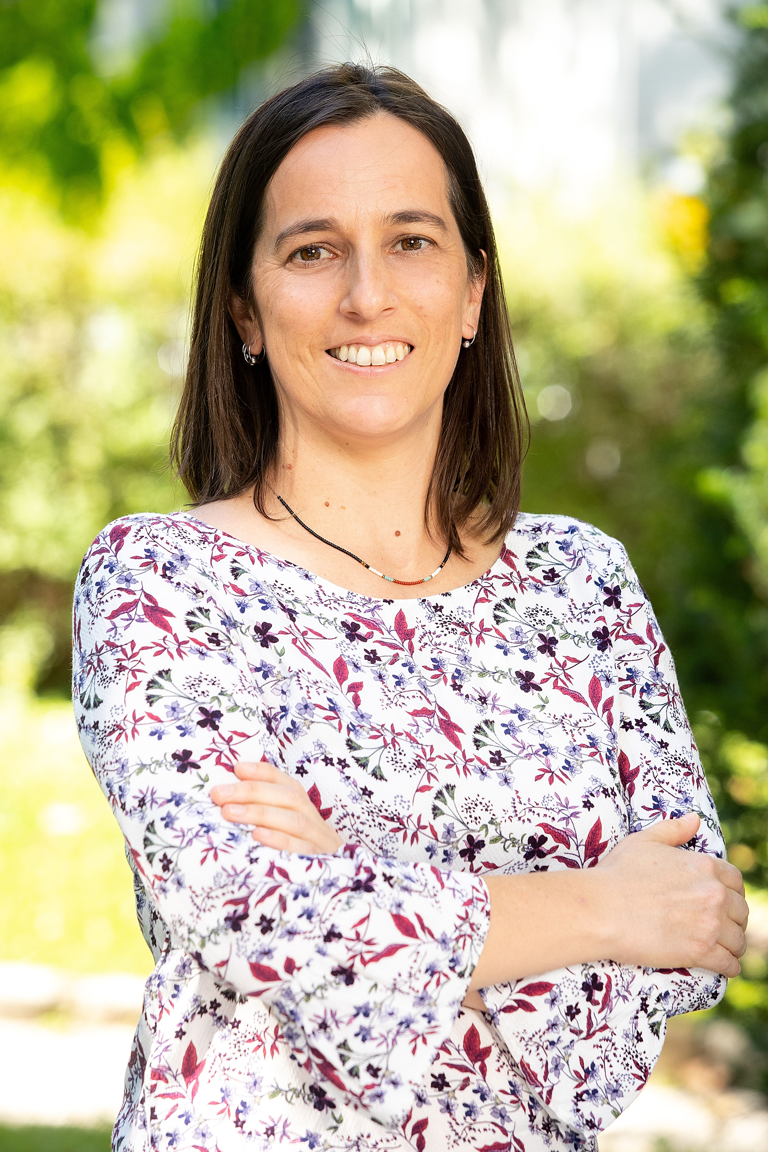
Children with certain malignant brain tumors that return—that is, recur—after treatment have a poor prognosis. The earlier the recurrence is detected, the better the chances of successful treatment. The research team led by Sibylle Madlener aims to identify characteristic features and find combinations with new, minimally invasive diagnostic and treatment methods that can be used for early diagnosis or to monitor treatment in children with brain tumors.
What are you investigating in your study?
We are looking for biomarkers—that is, specific cellular components of tumors found in bodily fluids—in young cancer patients in order to quickly detect recurrent tumors in children. In our study, “Identification of new CSF biomarkers and evaluation in recurrent MB and ATRT patients,” we are examining various tumor types for specific characteristics.
What are these characteristics?
Specifically, we were able to identify two biomarkers: microRNA clusters and cell-free DNA, or cell-free tumor DNA. MicroRNAs (miRNAs) are short, highly conserved, non-coding RNAs that play an important role in gene regulation. Cell-free DNA is a DNA structure that is randomly released into bodily fluids from surrounding cells due to cell death or other cellular reorganizations. This DNA often carries mutations that are frequently found in specific tumors.
What results did your research on the new biomarkers yield?
For the tumor types MB (medulloblastoma) and ETMR (embryonal tumor with multilayer rosettes), we were able to detect specific microRNA clusters in the blood and/or cerebrospinal fluid thanks to a new miRNA screening technology. In the case of MB, which occurs very frequently, we were not only able to identify the miRNA marker in patients’ serum but also detect it repeatedly as the disease progressed.
The miRNA markers were even more significant in ETMR patients. Here, we were able to detect the specific miRNAs in the patients’ cerebrospinal fluid without exception and with high statistical significance compared to other controls and tumor types. For this very rare and highly aggressive tumor type, we also observed a correlation between surgical removal followed by chemotherapy and the miRNA markers in the blood.
Together with our collaboration partners from the University of Freiburg in Germany and the University of Applied Sciences Vorarlberg, we are working on a novel method that enables rapid and simple detection of these specific miRNAs in patients’ blood—a so-called “lab-on-a-chip.” We were already able to publish our initial results last year in the highly prestigious journal *Advanced Materials*.
In addition to the promising miRNAs, we have also analyzed cell-free DNA—specifically, cell-free tumor DNA—from gliomas, which are brain tumors of the central nervous system. Using a highly sensitive droplet PCR method, we were able to detect a common mutation in the cerebrospinal fluid and plasma of patients with the extremely aggressive DIPG form of glioma. We were also able to detect duplications—known as amplifications—of individual gene segments, such as the MYC gene, by analyzing cell-free DNA in MB tumors. This MYC amplification fuels the growth and, subsequently, the rapid spread of the tumor. Such markers in cell-free DNA usually also indicate a poor prognosis for the course of the disease. That is why it is all the more important to detect them quickly and at an early stage.
What is the advantage of analyzing bodily fluids over examining tumor tissue?
The conventional method for obtaining a tissue sample is a biopsy, which usually involves surgery under general anesthesia. A relatively new procedure, known as a liquid biopsy, makes it possible to detect specific biomarkers that tumor cells release into bodily fluids such as blood or cerebrospinal fluid (CSF). This method is fast, reliable, virtually unlimited in scope, and minimally invasive for patients, as it eliminates the need for one or more complex surgeries. We have now, for the first time, identified biomarkers in blood serum and cerebrospinal fluid, enabling the rapid and simple detection of tumor cells.
The analysis of markers in bodily fluids will play a key role in the future in monitoring treatment and in prognosis for brain tumors.
About Sibylle Madlener
Sibylle Madlener is the principal investigator (PI) and lab manager of the Molecular Neuro-Oncology Laboratory at the University Clinic for Pediatrics and Adolescent Medicine at MedUni Vienna and Vienna General Hospital (AKH). She studied biology at the University of Innsbruck and moved to Vienna during her second cycle of studies to pursue a degree in human biology with a focus on molecular and tumor biology at the University of Vienna. She completed her master’s thesis in 2004 at the Clinical Institute of Laboratory Medicine at MedUni Vienna. From 2004 to 2009, she pursued her doctoral studies at the Institute of Clinical Pathology at MedUni Vienna, followed by a postdoctoral position at the University Clinic for Pediatrics and Adolescent Medicine at MedUni Vienna and Vienna General Hospital (AKH). She is the author and co-author of numerous publications and has already secured national and international research grants.

Chronic lymphocytic leukemia (CLL) is the most common blood cancer in the Western world, affecting primarily older adults. Although the disease can be treated very effectively today, it cannot yet be cured. In his study, Rainer Hubmann examines the origins of CLL and whether a mutation in a specific gene is responsible for its development. These findings could be crucial for advances in diagnosis and treatment.
What are you investigating in your study?
Our study, “Characterization of NOTCH2 gain-of-function N2ΔEC deletions as a possible causal genetic lesion in CLL,” examines whether a specific mutation—the NOTCH2 mutation—leads to the development of chronic lymphocytic leukemia. Using tumor samples collected from approximately 100 patients over the past 10 years, we are investigating whether these NOTCH2 mutations are detectable in all patients and in all leukemia cells. Only if the mutations are present in all of these cells and are directly linked to the spread of CLL cells are they considered disease-defining.
What is a NOTCH2 mutation?
NOTCH2 is responsible for the immortality of stem cells: As long as the stem cells remain in the stem cell niche, they produce new body cells through division, which can mature into specific tissue cells. Since NOTCH2 is a so-called stem cell factor, it could play a decisive role in the development of leukemia as a cancer-causing oncogene.
The precursor cells of CLL are a subtype of B cells, which are white blood cells that play a crucial role in the immune system. The stem cell niche of these B cells is located in the spleen. It appears that a NOTCH2 mutation occurs in one of these B cells there, allowing it to proliferate uncontrollably as a CLL cell.
What results has your study yielded so far?
We have succeeded in demonstrating that the alteration of NOTCH2 in leukemia cells occurs at the mRNA level through the loss of a specific segment of NOTCH2 (NOTCH2ΔNRR). This happens through “aberrant splicing,” a process in which specific sequence segments are selectively excised from a precursor mRNA. We were able to demonstrate that the most common loss of NOTCH2ΔNRR is associated with a congenital genetic variation. This variation is part of an evolutionarily ancient NOTCH2 gene variant, which the research team named NOTCH2*1A1. Individuals with this congenital NOTCH2*1A1 gene variant have a predisposition to CLL and other NOTCH2-associated tumors. The initial genetic alteration is likely a double-strand DNA break, during which the repair mechanism likely results in the exchange of genetic information between two NOTCH2 gene variants. Based on this finding, we were able to develop a simple genetic test that can be performed by any laboratory and for which a patent application has already been filed.
The results of our project are thus expected not only to explain the emergence of the NOTCH2 subtype in CLL cells but also have the potential to answer key open questions regarding geographic distribution, hereditary factors, and sex-specific differences in the development of certain leukemias and tumors.
What do these findings mean for the treatment and diagnosis of CLL?
If it can be demonstrated that the NOTCH2 gene mutation is a disease-defining event, this would not only represent a breakthrough in our understanding of the development of CLL, but it could also lead to the development of targeted drugs against the NOTCH2 mutation that might have the potential to cure CLL. In terms of diagnosis and prognosis, the presence of the mutation could indicate the onset of CLL. Following treatment, detection of the mutation would suggest that a recurrence of the disease is likely. In both cases, the disease could thus be detected and treated at an early stage.
About the Author
Rainer Hubmann studied biology and genetics at the University of Vienna. After graduating in 1996, he began his PhD program at MedUni Vienna, which he successfully completed in 2002. This was followed by postdoctoral positions at MedUni Vienna and the Ludwig Boltzmann Cluster Oncology. Since 2011, he has been a Principal Investigator at MedUni Vienna and the Comprehensive Cancer Center (CCC). Hubmann is the author and co-author of numerous publications, has already secured several awards and grants, is active in teaching, and holds three patents. In addition, he is a member of the Austrian Society for Hematology and Medical Oncology, as well as a member of the Comprehensive Cancer Center (CCC) at MedUni Vienna and the Vienna General Hospital (AKH).
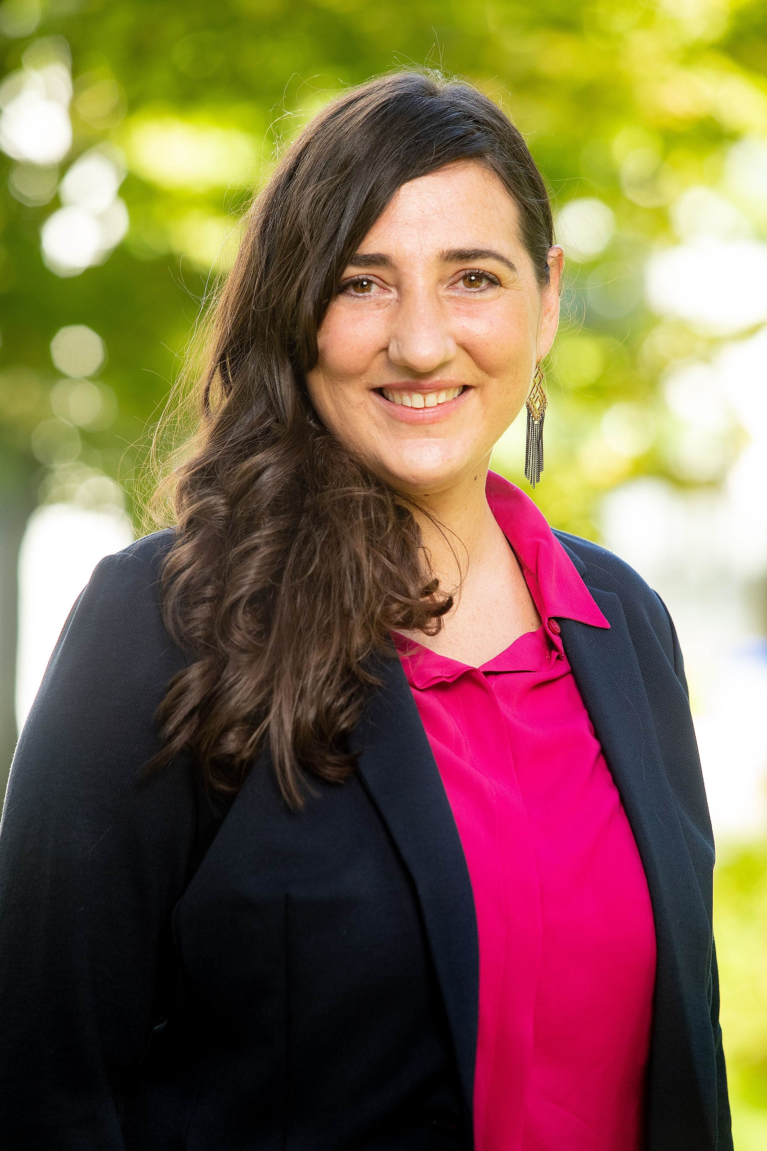
Each year, approximately 1,600 people are diagnosed with skin cancer. Although there are already numerous treatment approaches for metastatic skin cancer, they are not always effective because patients develop resistance to the therapy. Verena Paulitschke’s research aims to better understand resistance in the treatment of skin cancer and to detect it at an early stage.
What are you researching in your studies?
Our studies focus on metastatic skin cancer and why some patients develop resistance to treatments. Over the past ten years, groundbreaking and effective therapeutic approaches have been developed for treating this disease, both through targeted therapy directed at the tumor cells and through the activation of the immune system. Despite a good initial response, resistance to targeted therapy develops on average after about 15 months, or only about half of patients benefit from immunotherapy. One possible explanation is that melanomas are highly adaptable, which allows tumor cells to interfere with biological processes. We have therefore set out to investigate these various resistance mechanisms in order to better understand them and to generate proposals on how to overcome them or detect them early.
The first study focused on resistance mechanisms to targeted therapy and was published in 2019 in the EMBO Journal under the title “Proteomic identification of a marker signature for MAPKi resistance in melanoma.” In this study, we characterized, among other things, two proteins associated with response to therapy and also patented a corresponding marker signature. This work was honored with the 2019 Fleur-Hiege Prize.
In our ongoing study, “Identification of novel markers to predict response to anti-PD-1 immunotherapy in melanoma applying proteomics,” we have been searching for markers that could predict response to immunotherapy at an early stage—so-called predictive markers. This study also serves as the PhD thesis of my student Nina Zila, the first author of this paper.
How are you investigating this resistance?
Using proteomics, we analyze serum samples and tumor cell cultures from melanoma patients who have been treated with immunotherapy, and we examine the molecular mechanisms responsible for whether patients respond to therapy or not. We then conduct functional experiments and clinical validations to substantiate the results. It is particularly important here to be able to use well-characterized patient samples. To this end, we were able to draw on the biobank at the University Hospital of Zurich, led by Prof. Levesque and Prof. Dummer, as well as our own biobank in the Department of Dermatology, led by Prof. Höller, Prof. Stary, and Prof. Weninger.
What is proteomics?
The proteome consists of all proteins present in a cell or an organism under defined conditions and at a specific point in time. Proteomics examines this proteome using biochemical methods and provides comprehensive and biologically relevant insight into cellular processes. This allows us to identify how and under what circumstances proteins change in a way that leads to resistance.
These analyses require highly sensitive equipment, and we had the samples analyzed at ETH Zurich by Prof. Aebersold at the Institute of Molecular Systems Biology and at the University of Vienna by Prof. Gerner at the Institute of Analytical Chemistry.
What findings have you obtained in your ongoing study?
It is particularly interesting to us that we can observe mechanisms in serum and tumor cells similar to those seen with targeted therapy. The tumor cells appear to change in such a way that they grow and metastasize less vigorously. Therefore, a better understanding of these processes could be beneficial for both targeted therapy and immunotherapy.
Thanks to the markers in the tumor cell proteome, we were able to identify a distinct immunophenotype—that is, to detect specific antigens on the tumor cells that are responsible for resistance and play a potential role in the tumor microenvironment.
We were also able to identify promising markers in serum and validate them using various methods, so that we now have both a tumor and a serum marker signature that we hope will help us predict resistance in the future. If this is possible, it would allow us to determine early on whether a patient will benefit from immunotherapy, thereby helping to ensure that they receive the best possible treatment without losing valuable treatment time.
About the Author
Verena Paulitschke is a senior physician in dermatology at the Department of Dermatology at the Medical University of Vienna. Her research focuses on basic dermato-oncological research and translational research. She studied medicine at MedUni Vienna and also completed her residency, PhD, and habilitation at the University Clinic for Dermatology there. She completed a two-year research fellowship at ETH Zurich and the Department of Dermatology at the University Hospital of Zurich. She has been an associate professor since 2018. Her research focuses on evaluating resistance mechanisms to targeted therapies and immunotherapy and identifying predictive markers in metastatic melanoma. She has already won numerous awards for her publications.