
The MIC nodes invite you to take part and interact actively in a virtual poster tour through the most innovative and exciting topics related to medical imaging presented by young scientist.
Postersessions at a glance
MIC Nodes | Postersessions 14:00 - 14:45
ED-B Fibronectin as a potential marker for tumour microenvironment and fibrosis
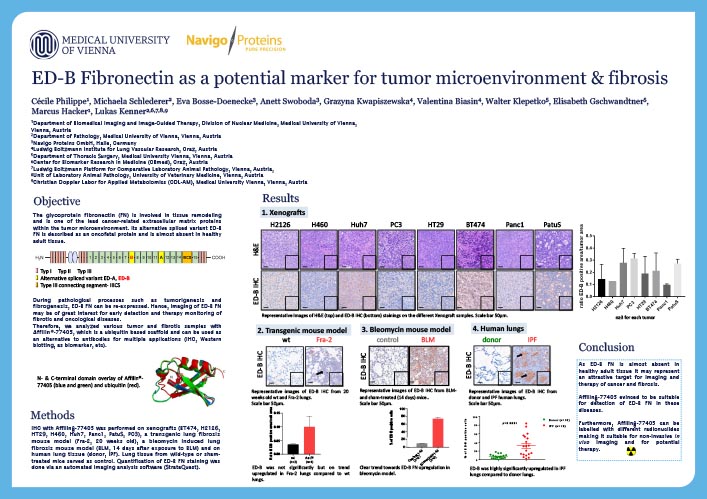
Cecile Philippe
Department of Biomedical Imaging and Image-Guided Therapy, Division of Nuclear Medicine, Medical University of Vienna
Introduction
The glycoprotein fibronectin (FN) is involved in tissue remodelling and is one of the lead cancer-related extracellular matrix proteins within the tumour microenvironment. Its alternative spliced variant ED-B FN is described as an oncofetal protein and is almost absent in healthy adult tissue. During pathological processes such as tumorigenesis and fibrogenesis, ED-B FN can be re-expressed. Hence, imaging of ED-B FN may be of great interest for early detection and therapy monitoring of fibrotic and oncological diseases. Therefore, we analysed various tumour and fibrotic samples with Affilin®-77405, which has been developed as a potential biomarker for ED-B FN.
Development of small molecule PET-tracers targeting PD-L1
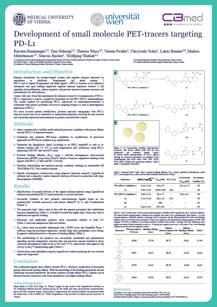
Karsten Bamminger
Department of Biomedical Imaging and Image-guided Therapy, Division of Nuclear Medicine, Medical University of Vienna, Vienna, Austria
CBmed GmbH - Center for Biomarker Research in Medicine, Graz, Austria
Introduction
Immune checkpoints are receptor-ligand systems and regulate immune responses by stimulation or inhibition. Programmed cell death protein 1 (PD-1) and its ligand Programmed cell death ligand 1 (PD-L1) function as an inhibitory checkpoint and upon binding negatively regulate immune responses, obstruct T cell signaling and proliferation, induce apoptosis and prevent excessive immune reactions and autoimmunity for self-tolerance. Cancer cells may abuse this mechanism for immune evasion by overexpression of PD-L1. PD-L1 expression is used as a predictive biomarker for anti-PD-1/PD-L1 immunotherapy.
The current method for quantifying PD-L1 expression by immunohistochemistry is confronted with intrinsic problems of invasive sampling (biopsy) as well as heterogeneous expression of PD-L1.
For more accurate patient stratification, positron emission tomography with PD-L1 selective tracers may be an alternative to immunohistochemistry allowing for non-invasive and concurrent expression measurements in primary and metastatic tumors.
Hydrobenzoin esters of arecaidine as potential PET tracers for muscarinic acetylcholine receptors: synthesis, characterization and docking experiments.
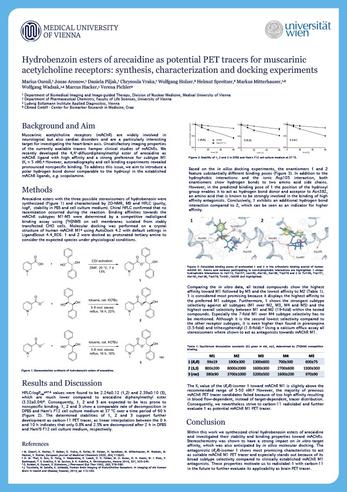
Marius Ozenil
Department of Biomedical Imaging and Image-guided Therapy, Division of Nuclear Medicine, Medical University of Vienna
Introduction
Muscarinic acetylcholine receptors (mAChR) are widely involved in neurological but also cardiac disorders and are a particularly interesting target for investigating the heart-brain axis. Unsatisfactory imaging properties of the currently available tracers hamper clinical studies of mAChRs. We recently developed the 4,4’-difluordiphenylmethyl ester of arecaidine as mAChR ligand with high affinity and a strong preference for subtype M1 (Ki = 5 nM). However, autoradiography and cell binding experiments revealed pronounced nonspecific binding. To address this issue, we aim to introduce a polar hydrogen bond donor comparable to the hydroxyl in the established mAChR ligands, e.g. scopolamine.
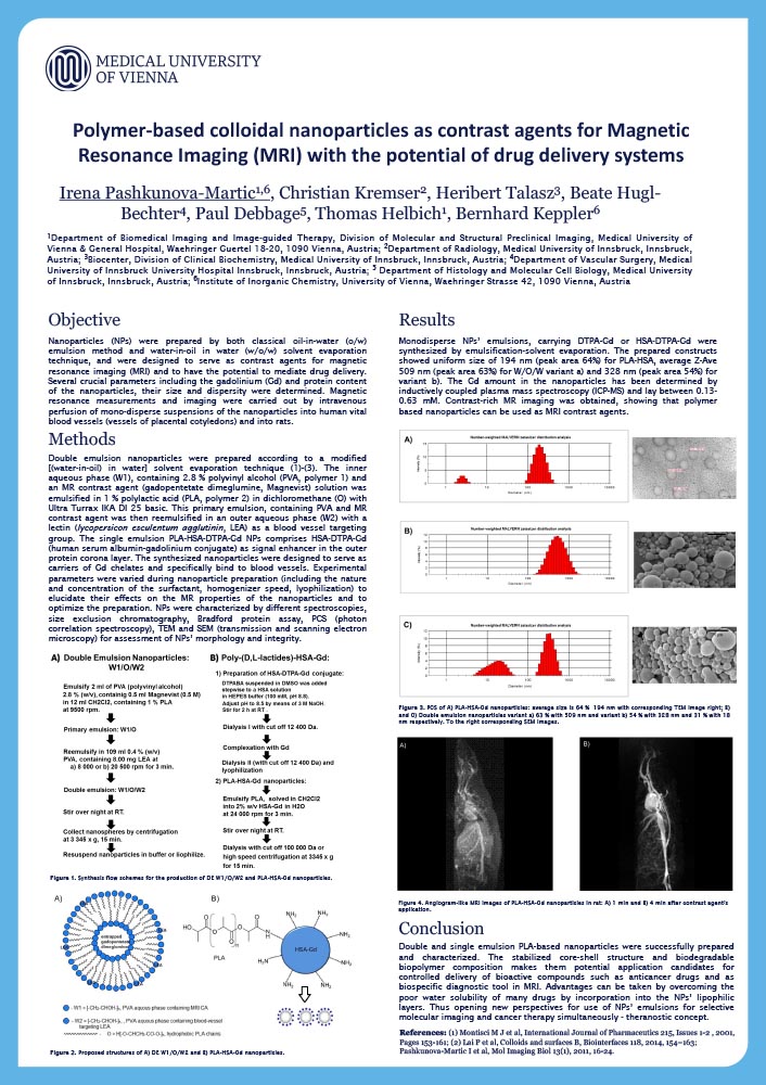
Polymer-based colloidal nanoparticles as contrast agents for Magnetic Resonance Imaging (MRI) with the potential of drug delivery systems
Irena Pashkunova-Martic
Department of Biomedical Imaging and Image-guided Therapy, Division of Molecular and Structural Preclinical Imaging, Medical University of Vienna
Institute of Inorganic Chemistry, University of Vienna
Introduction
Nanoparticles (NPs) were prepared by both classical oil-in-water (o/w) emulsion method and water-in-oil in water (w/o/w) solvent evaporation technique, and were designed to serve as contrast agents for magnetic resonance imaging (MRI) and to have the potential to mediate drug delivery. Several crucial parameters including the gadolinium (Gd) and protein content of the nanoparticles, their size and dispersity were determined. Magnetic resonance measurements and imaging were carried out by intravenous perfusion of mono-disperse suspensions of the nanoparticles into human vital blood vessels (vessels of placental cotyledons) and into rats.
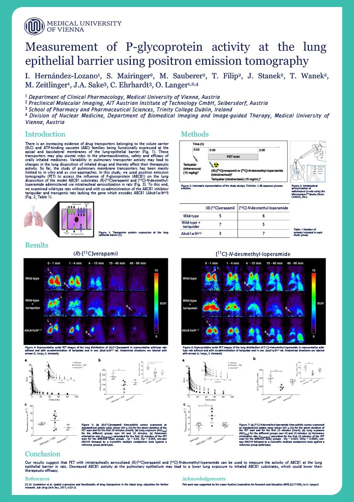
Measurement of P-glycoprotein activity at the lung epithelial barrier using positron emission tomography
Irene Hernández Lozano
Department of Clinical Pharmacology, Medical University of Vienna
Introduction
Drug transporters belonging to the solute carrier (SLC) and ATP-binding cassette (ABC) families are functionally expressed at the apical (airway-facing) and basolateral (blood-facing) membranes of the lung epithelial barrier. These transporters might play pivotal roles in the pharmacokinetics, safety and efficacy of orally inhaled medicines. Variability in pulmonary transporter activity may lead to changes in the lung disposition of inhaled drugs and thereby contribute to interindividual variability in therapeutic response. So far, the study of pulmonary transporters has been mainly limited to in vitro and ex vivo (e.g. isolated perfused rodent lungs) approaches. Inhibition of P-glycoprotein (ABCB1), which is localized to the apical membrane of the lung epithelium, was shown to increase the absorption of inhaled model ABCB1 substrates from the lungs into the systemic circulation.
In this study, we used positron emission tomography (PET) to assess the influence of ABCB1 on the lung disposition of the model ABCB1 substrate (R)-[11C]verapamil in rats. PET scans were carried out after intratracheal aerosolisation of (R)-[11C]verapamil in three Sprague Dawley rat groups: wild-type as control, Abcb1a/b(-/-) rats, and wild-type with co-administration of the ABCB1 inhibitor tariquidar, which was administered intravenously (15 mg/kg) and intratracheally (15 mg/ml). Lung exposure (AUClung) to (R)-[11C]verapamil was significantly lower (p<0.05) in tariquidar-treated rats (58±37 min) and in Abcb1a/b(-/-) rats (67±24 min) compared to untreated wild-type rats (132±22 min), which demonstrated an involvement of ABCB1 in the pulmonary absorption of (R)-[11C]verapamil. Our results suggest that PET with intratracheally aerosolised (R)-[11C]verapamil can be used to measure the activity of ABCB1 at the lung epithelial barrier. Decreased ABCB1 activity at the pulmonary epithelium may lead to a lower lung exposure to inhaled ABCB1 substrates, which may lower their therapeutic efficacy.
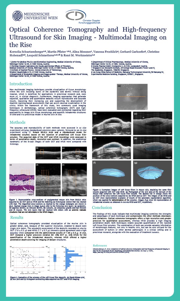
Optical Coherence Tomography and High-frequency Ultrasound for Skin Imaging-Multimodal Imaging on the Rise
Kornelia Schuetzenberger
Center for Medical Physics and Biomedical Engineering, Medical University of Vienna
Christian Doppler Laboratory for Ocular and Dermal Effects of Thiomers
Introduction
New multimodal imaging techniques enabling the visualization of tissue structures within the epidermis and dermis non-invasively are favorable for both preclinical research and clinical diagnosis. Furthermore, imaging approaches that generate objective, qualitative and quantitative datasets ensure reproducible and accurate results. Within the framework of this study, the two frequently used imaging techniques in dermatology, optical coherence tomography (OCT) and high-frequency ultrasound (HFUS), have been assessed and compared concerning their applicability to image skin tissue morphology and quantify intradermal structures in murine skin.
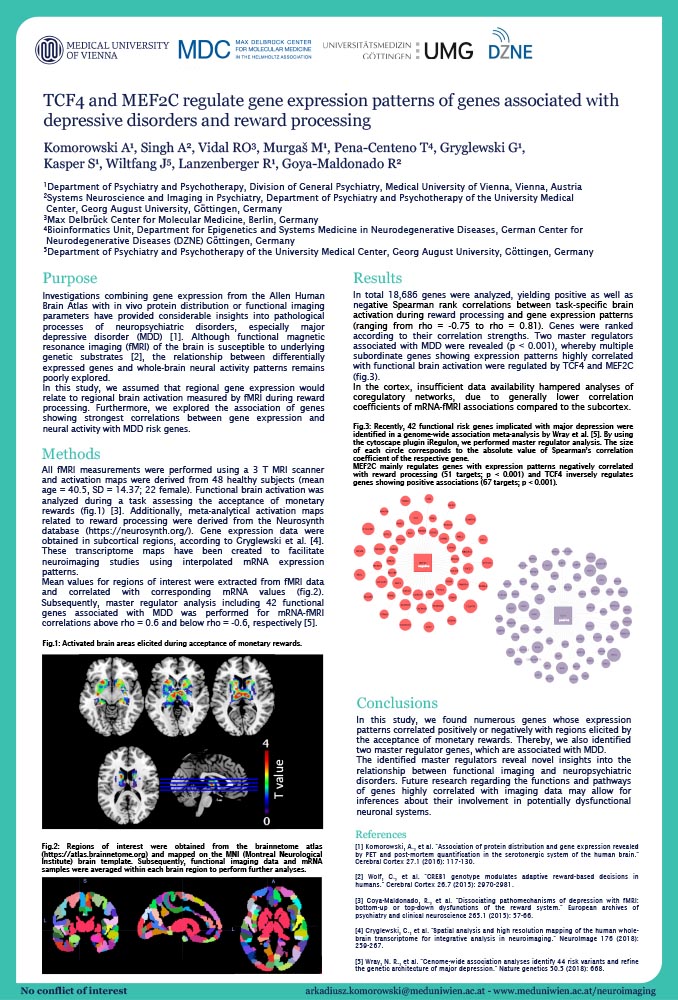
TCF4 and MEF2C regulate gene expression patterns of genes associated with depressive disorders and reward processing
Arkadiusz Komorowski
Department of Psychiatry and Psychotherapy, Division of General Psychiatry, Medical University of Vienna
Introduction
Investigations combining gene expression from the Allen Human Brain Atlas with in vivo protein distribution or functional imaging parameters have provided considerable insights into pathological processes of neuropsychiatric disorders, especially major depressive disorder (MDD) [1]. Although functional magnetic resonance imaging (fMRI) of the brain is susceptible to underlying genetic substrates [2], the relationship between differentially expressed genes and whole-brain neural activity patterns remains poorly explored.
In this study, we assumed that regional gene expression would relate to regional brain activation measured by fMRI during reward processing. Furthermore, we explored the association of genes showing strongest correlations between gene expression and neural activity with MDD risk genes.
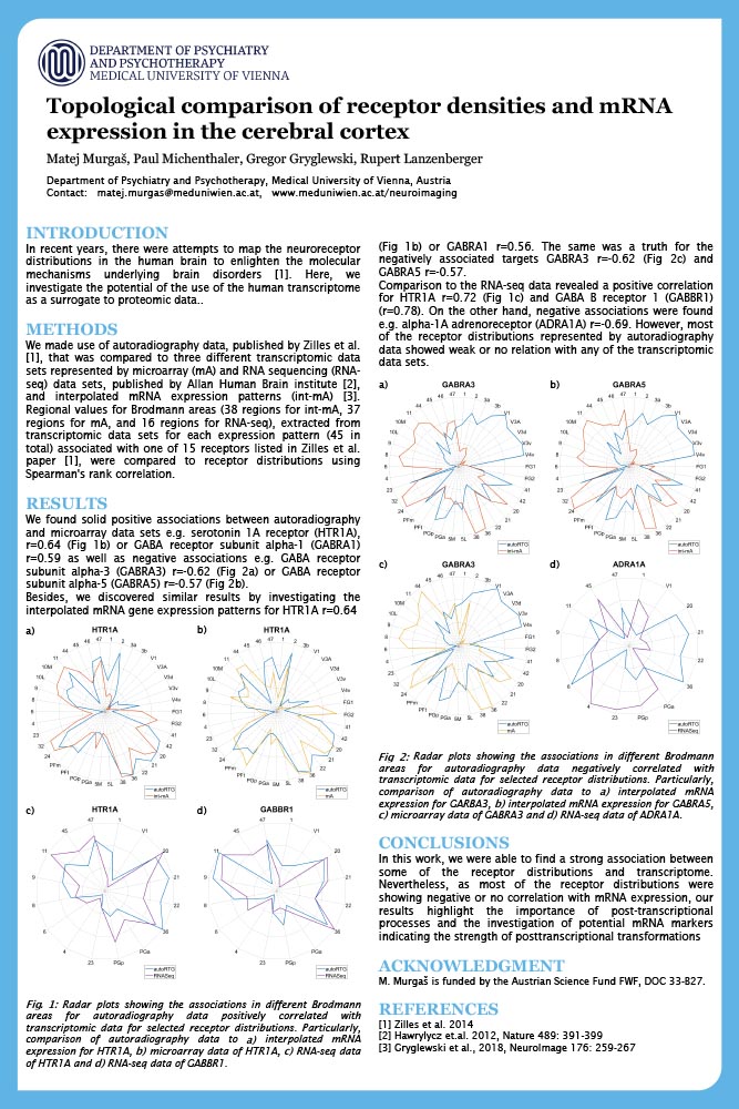
Topological Comparison of Receptor Densities and mRNA Expression in the Cerebral Cortex
Matej Murgaš
Department of Psychiatry and Psychotherapy, Medical University of Vienna
Introduction
In recent years, there were attempts to map the neuroreceptor distributions in the human brain to enlighten the molecular mechanisms underlying brain disorders. Here, we investigate the potential of the use of the human transcriptome as a surrogate to proteomic data.
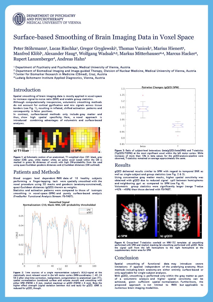
Surface-based Smoothing of Brain Imaging Data in Voxel Space
Peter Stöhrmann
Department of Psychiatry and Psychotherapy, Medical University of Vienna
Introduction
Spatial smoothing of brain imaging data is mostly applied in voxel-space to increase signal-to-noise ratio (SNR) and enable group statistics. Although computationally inexpensive, volumetric smoothing methods do not account for cortical gyrification and mix signals across tissue borders (see Fig. 1), resulting in inflated, shifted activation patterns and consequently in false positives.
In contrast, surface-based methods only include gray matter and thus, show high spatial specificity. Here, a novel approach is introduced combining advantages of volumetric and surface-based analyses.
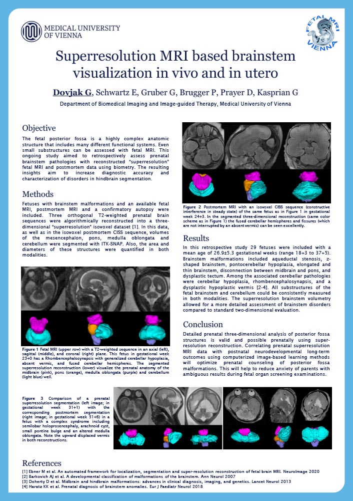
Superresolution MRI based brainstem visualization in vivo and in utero
Gregor O Dovjak
Department of Biomedical Imaging and Image-guided Therapy, Medical University of Vienna
Introduction
The fetal posterior fossa is a highly complex anatomic structure that includes many different functional systems. Even small substructures can be assessed with fetal MRI. This ongoing study aimed to retrospectively assess prenatal brainstem pathologies with reconstructed “superresolution” fetal MRI and postmortem data using biometry. The resulting insights aim to increase diagnostic accuracy and characterization of disorders in hindbrain segmentation.
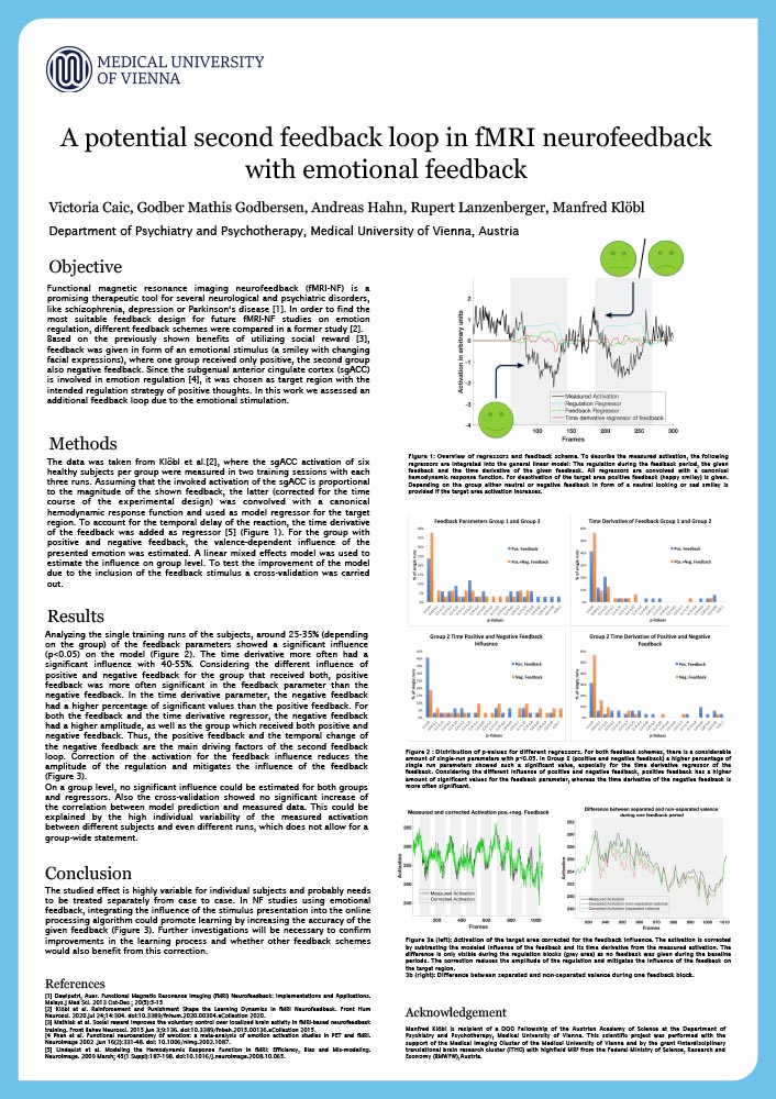
A potential second feedback loop in fMRI neurofeedback with emotional feedback
Victoria Caic
Department of Psychiatry and Psychotherapy, Medical University of Vienna
Introduction
Functional MRI neurofeedback (fMRI-NF) is a promising therapeutic tool for several neurological and psychiatric disorders. In order to find the most suitable feedback design for future fMRI-NF studies on emotion regulation, different feedback schemes were compared [1]. Based on the previously shown benefits of utilizing social reward [2], feedback was given in form of an emotional stimulus (a smiley with changing facial expressions). One group received only positive feedback, the second group received also negative feedback. Since the target region, the subgenual anterior cingulate cortex (sgACC), is involved in emotional processing [3], an additional feedback loop due to emotional stimulation would be possible. The size and significance of this effect were estimated in this work.
Neuroplastic effects of SSRIs evaluated with learning tasks and fMRI

Murray B Reed
Department of Psychiatry and Psychotherapy, Medical University of Vienna
Introduction
One core function of cognitive flexibility is the use of past memory to adapt to environmental changes. Serotonin has been implicated in executive, learning and affective functions and selective serotonin reuptake inhibitors (SSRIs) are the current first-line treatment for mood and anxiety disorders. Despite recent progress in elucidating the relationships of the serotonergic system and their effects on learning and retrieval of information, far from all is understood.
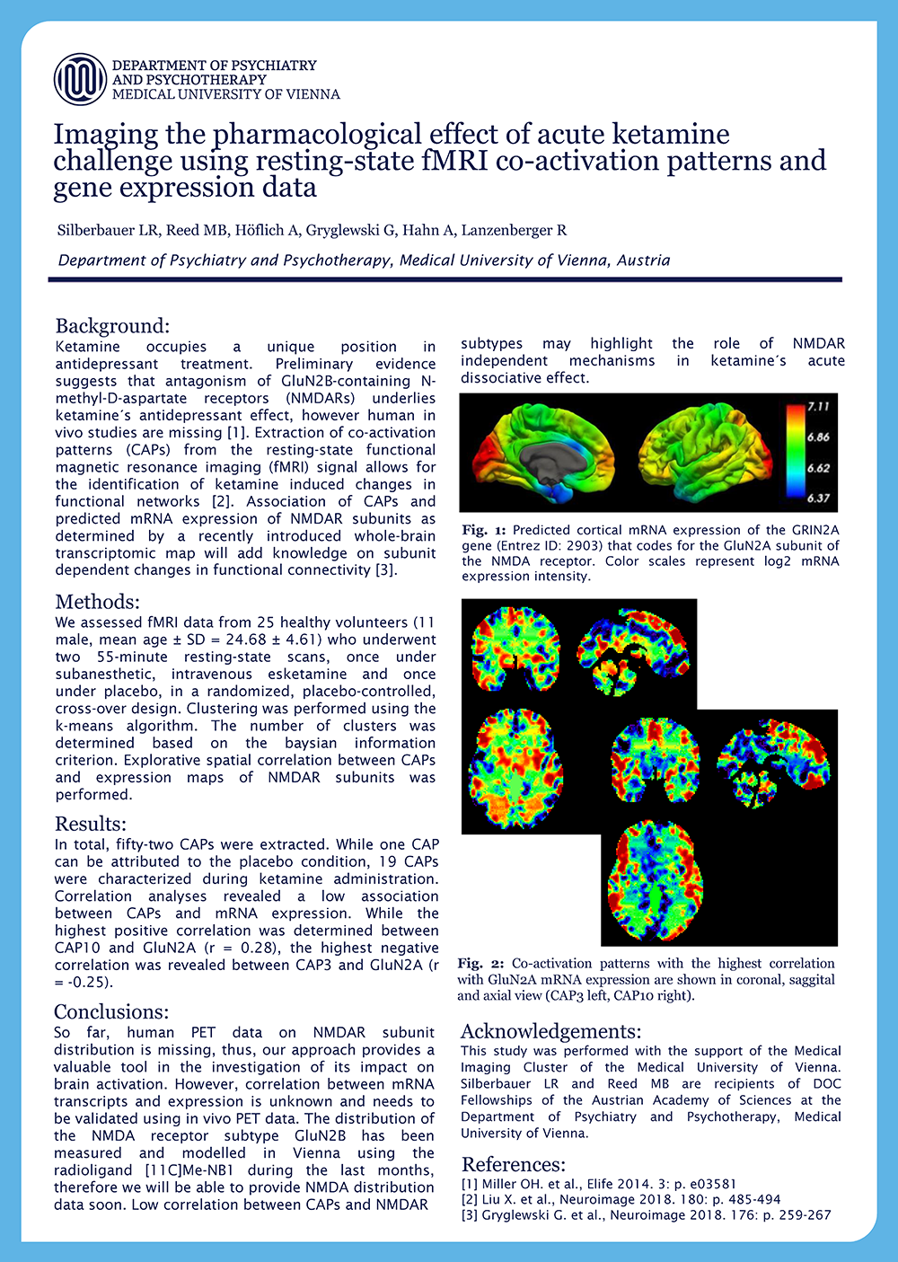
Imaging the pharmacological effect of acute ketamine challenge using resting-state fMRI co-activation patterns and gene expression data
Leo R Silberbauer
Department of Psychiatry and Psychotherapy, Medical University of Vienna
Introduction
Ketamine occupies a unique position in antidepressant treatment. Preliminary evidence suggests that antagonism of GluN2B-containing N-methyl-D-aspartate receptors (NMDARs) underlies ketamine´s antidepressant effect, however human in vivo studies are missing [1]. Extraction of co-activation patterns (CAPs) from the resting-state functional magnetic resonance imaging (fMRI) signal allows for the identification of ketamine induced changes in functional networks [2]. Association of CAPs and predicted mRNA expression of NMDAR subunits as determined by a recently introduced whole-brain transcriptomic map will add knowledge on subunit dependent changes in functional connectivity [3].
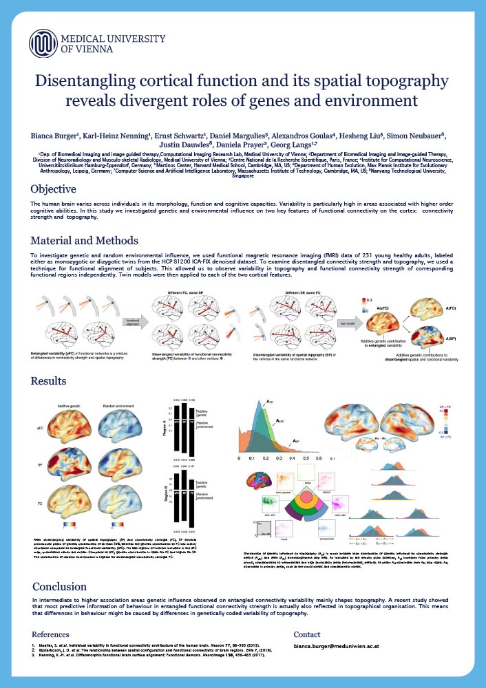
Disentangling cortical function and its spatial topography reveals divergent roles of genes and learning
Bianca Burger
Department of Biomedical Imaging and image guided therapy, Computational Imaging Research Lab, Medical University of Vienna
Introduction
The human brain varies across individuals in its morphology, function and cognitive capacities. Variability is particularly high in areas associated with higher order cognitive abilities. In this study we investigated genetic and environmental influence on two key features of functional connectivity on the cortex: connectivity strength and topography.
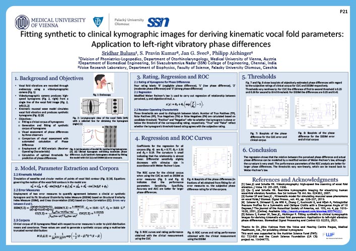
Fitting synthetic to clinical kymographic images for deriving kinematic vocal fold parameters: Application to left-right vibratory phase differences
Philipp Aichinger
Division of Phoniatrics-Logopedics, Department of Otorhinolaryngology, Medical University of Vienna
Introduction
In this paper we present the extraction of kinematic vocal fold parameters from videokymographic images, the visual estimation of phase differences between the left and right vocal folds and the training of a convolutional neural network for parameter extraction from synthetic kymograms. We used a model of vocal fold vibrations with kinematic parameters to generate synthetic kymograms and propose a fitting procedure for error minimization for extracting kinematic parameters from the images. By using the “Structural Dissimilarity Index Measure” (DSSIM) and the “Cross Uncorrelation” (CUC) as error measures, the minimization procedure was used to evaluate 55 clinical kymograms and two sets of 50 synthetic kymograms each. Fitting the clinical kymograms yielded probability density functions (PDFs) of the model parameters and their correlation coefficients, reflecting a multivariate normal distribution.
These distributions were used to generate the synthetic corpora. The relative parameter estimation errors range up to 15% for the most significant parameters, indicating acceptable performance of the fitting procedure. Additionally, the phase difference was assessed by three observers with integer ratings ranging from 0 (negligible) to 3 (large phase difference). In order to quantify the agreement among the observers, Fleiss’ Kappa and its Standard Error were calculated. ROC analysis was carried out for estimating thresholds for predicting the ratings. Performance measures such as Sensitivity, Specificity and Accuracy were calculated. Moderate to good (0.69 to 0.91) accuracies for the clinical corpus and very good accuracy (0.92 to 1.00) for the synthetic corpora were observed.
Finally, the convolutional neural networks (CNN) were trained to perform extraction of vocal fold parameters from a synthetic corpus of ten-thousand kymograms. Fitting kymograms with the model typically takes one hour per kymogram as compared to less than a minute with the CNN. Moreover, the error of the CNN regression is less for all parameters except for the left and right phase. In the future, transfer learning will be applied to fine tune the CNNs with natural training data.
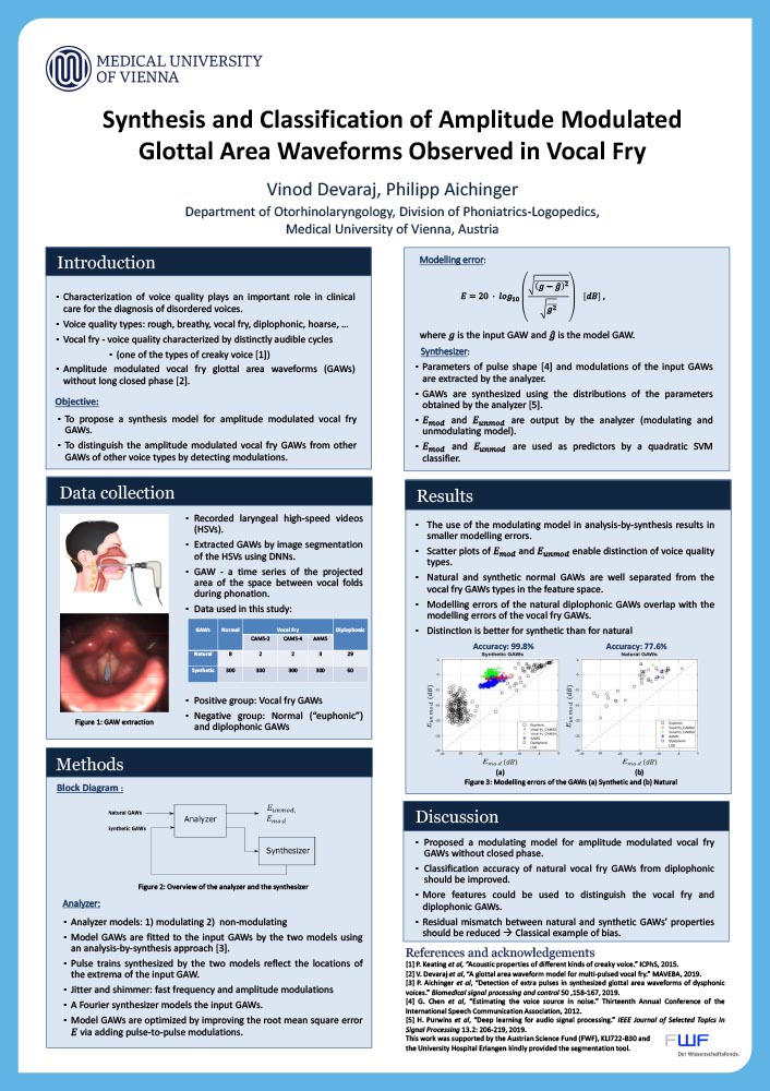
Synthesis and Classification of Amplitude Modulated Vocal Fry GAWs
Vinod Devaraj
Deptartment of Otorhinolaryngology, Division of Phoniatrics-Logopedics, Medical University of Vienna
Introduction
Dysphonia, i.e., the dysfunction of human voice, is often accompanied by voice sounds, the auditory qualities of which diverge from the norm. Characterization of voice qualities is important for the clinical care of disordered voices, because they aid the indication, selection, evaluation and optimization of clinical treatment techniques. Vocal fry is a voice quality that is also designated as creaky voice [1]. In this study, videos of the vocal folds during sustained phonation are obtained with an endoscopic laryngeal high-speed camera using a frame rate of 4000 frames per second while the audio files are recorded simultaneously. The voice samples are annotated by expert listeners with regard to presence or absence of vocal fry based on the auditory temporal segregation of individual glottal pulses. Glottal area waveforms (GAWs) are extracted from high-speed videos using a deep neural network (DNN) for graphical segmentation implemented in an available tool. A GAW is a time series of the glottal area, i.e., the projected area of the space between the vocal folds, which vibrate during phonation.
Traditionally, vocal fry is characterized by low frequencies and a long closed phase of the glottis.
However, we also observe amplitude modulated vocal fry GAWs without long closed phases (positive group). The negative group in this study consists of euphonic and diplophonic GAWs. GAWs are synthesized for the positive and the negative groups based on the parameter distribution of the original GAWs. Modulated and unmodulated GAWs are fitted to the input GAWs (synthesized) using an analysis and synthesis approach described in [2]. Modelling errors are obtained for modulated and unmodulated GAWs with the corresponding input GAW as reference. An SVM classifier with a quadratic kernel achieves a 5-fold cross validated accuracy of 93 % when using the two modelling errors as predictors. The accuracy drops by few percent when natural GAWs are included as input GAWs, which may be interpreted as a consequence of a residual mismatch of the natural and synthetic GAWs’ properties. Also, natural GAWs are dislocated from the respective voice type’s synthetic GAWs in the feature space. This should be addressed in the future by improving the parameter estimation of the original GAWs, and by improving the naturalness of the synthetic GAWs.
This work was supported by the Austrian Science Fund (FWF), KLI 722-B30 and the University Hospital Erlangen kindly provided the segmentation tool.
Dynamic memory to alleviate catastrophic forgetting in continuous learning settings
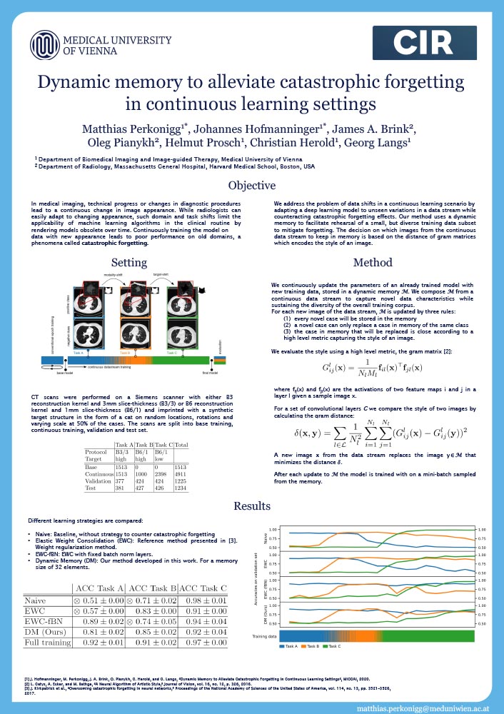
Matthias Perkonigg
Department of Biomedical Imaging and Image guided Therapy, Medical University of Vienna
Introduction
In medical imaging, technical progress or changes in diagnostic procedures lead to a continuous change in image appearance. While radiologists can easily adapt to changing appearance, such domain and task shifts limit the applicability of machine learning algorithms in the clinical routine by rendering models obsolete over time. Continuously training the model on data with new appearance leads to poor performance on old domains, a phenomena called catastrophic forgetting.
In this work, we address the problem of data shifts in a continuous learning scenario by adapting a deep learning model to unseen variations in a data stream while counteracting catastrophic forgetting effects. Our method uses a dynamic memory to facilitate rehearsal of a small, but diverse training data subset to mitigate forgetting. The decision on which images from the continuous data stream to keep in memory is based on the distance of gram matrices which encodes the style of an image.
We evaluated our approach on routine clinical CT data of the lung obtained with two different scanner protocols and synthetic classification tasks. Experiments show that dynamic memory counters catastrophic forgetting in a setting with multiple data shifts. Unlike current state of the art continuous learning methods, our approach operates without the necessity for explicit knowledge about when data shifts occur in the stream.
Identification of aggressive breast cancer lesions with [18F]-FDG-PET/CT in combination with machine learning and data pre-processing
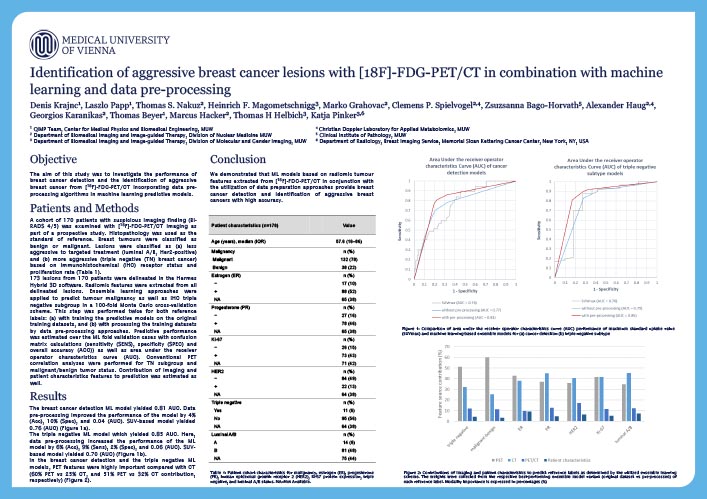
Denis Krajnc
QIMP Team, Center for Medical Physics and Biomedical Engineering, Medical University of Vienna
Introduction
The aim of this study was to investigate the performance of breast cancer detection and the identification of aggressive breast cancer from 18 F] FDG PET/CT incorporating data pre processing algorithms in machine learning predictive models.
Predictive modelling for prognostic stratification of head and neck cancer patients using multi-omics data
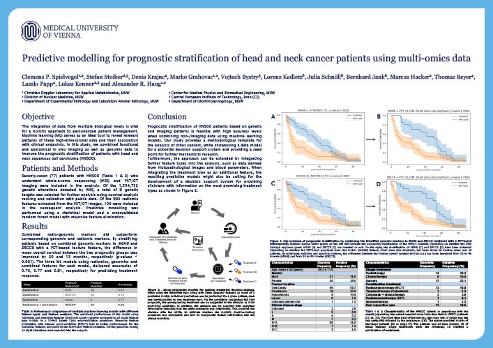
Clemens P Spielvogel
Christian Doppler Laboratory for Applied Metabolomics
Division of Nuclear Medicine, Medical University of Vienna
Introduction
The integration of data from multiple biological levels is vital for a holistic approach to personalized patient management. Here, machine learning (ML) serves as an ideal tool to reveal relevant patterns of these high-dimensional data and their association with clinical endpoints. In this study, we combined functional and anatomical in vivo imaging as well as genomic data to improve the prognostic stratification of patients with head and neck squamous cell carcinoma (HNSCC).
Voxel-wise Nuisance Regression for Suppressing the Residual Motionin Resting state in-utero fMRI
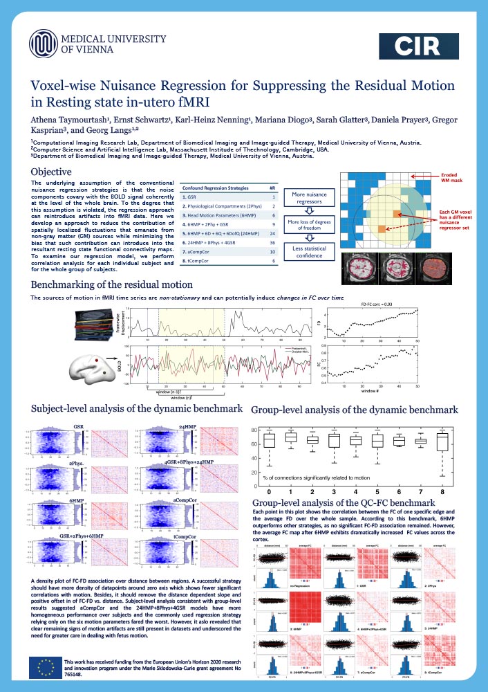
Athena Taymourtash
Computational Imaging Research Lab, Department of Biomedical Imaging and Image-guided Therapy, Medical University of Vienna
Introduction
The underlying assumption of the conventional nuisance regression strategies is that the noise components covary with the BOLD signal coherently at the level of the whole brain. To the degree that this assumption is violated, the regression approach can reintroduce artifacts into fMRI data. Here we develop an approach to reduce the contribution of spatially localized fluctuations that emanate from non-gray matter (GM) sources while minimizing the bias that such contribution can introduce into the resultant resting state functional connectivity maps. To examine our regression model, we perform correlation analysis for each individual subject and for the whole group of subjects.
Flexible multi-turn multi-gap coaxial RF coils (MTMG-CCs) for 3 and 7 Tesla MRI

Lena Nohava
Université Paris-Saclay, CEA, CNRS, Inserm, BioMaps, Orsay, France
Introduction
Close-fitting and size-optimized radio frequency (RF) coils for 3 and 7 T 1H-MRI have gained great interest in RF coil engineering, especially coaxial coils (CCs) as they are flexible, lightweight, robust against inter-element coupling and can be ideally fitted to the anatomy. We propose the design of flexible multi-turn multi-gap coaxial coils (MTMG-CCs) allowing for CC size optimization depending on the target anatomical application.
Optimization and miniaturization of Rx-only coaxial coil interfacing
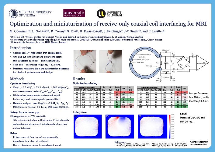
Michael Obermann
Division MR Physics, Center for Medical Physics and Biomedical Engineering, Medical University of Vienna
Introduction
RF coils made from coaxial cable were recently introduced. The cable’s inner and outer conductors are cut at positions opposite from each other and allow three separate currents to flow. A self-resonant structure is obtained without additional lumped components when the reactance presented by the two coaxial stubs cancels the loop’s inductance. In this work, the influence of the choice of interfacing components on the achieved SNR is studied on a coaxial coil.
Anatomy and physiology of the anterior eye segment

Rene Werkmeister
Center for Medical Physics and Biomedical Engineering, Medical University of Vienna
Christian Doppler Laboratory for Ocular Effects of Thiomers
Introduction
Non-invasive, non-contact imaging methods are essential for diagnostics and therapeutic interventions at the anterior eye segment. Due to the properties of the ocular tissues, optical modalities like optical coherence tomography (OCT) and confocal microscopy are ideally suited for visualizing the morphology of the anterior segment of healthy and diseased eyes with resolutions in the micrometer range. Advances in light source technology lead to a further improvement of the resolution allowing non-invasive imaging with almost cellular resolution.
Automatic segmentation and parameter estimation of the lower tear meniscus
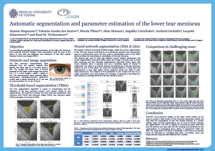
Hannes Stegmann
Center for Medical Physics and Biomedical Engineering, Medical University of Vienna
Christian Doppler Laboratory for Ocular Effects of Thiomers
Introduction
Dry eye disease (DED) is a prevalent and widespread multifactorial disease of the ocular surface that not only reduces the visual performance of the patient, but also has a measurable impact on its quality of life. The tear meniscus, which is the concave surface of tear fluid at the upper and lower eyelid margins, contains most of the tear fluid and has been widely studied in the context of DED.
Evaluating CNNs for image upsampling of anisotropic MRIs in radiation oncology
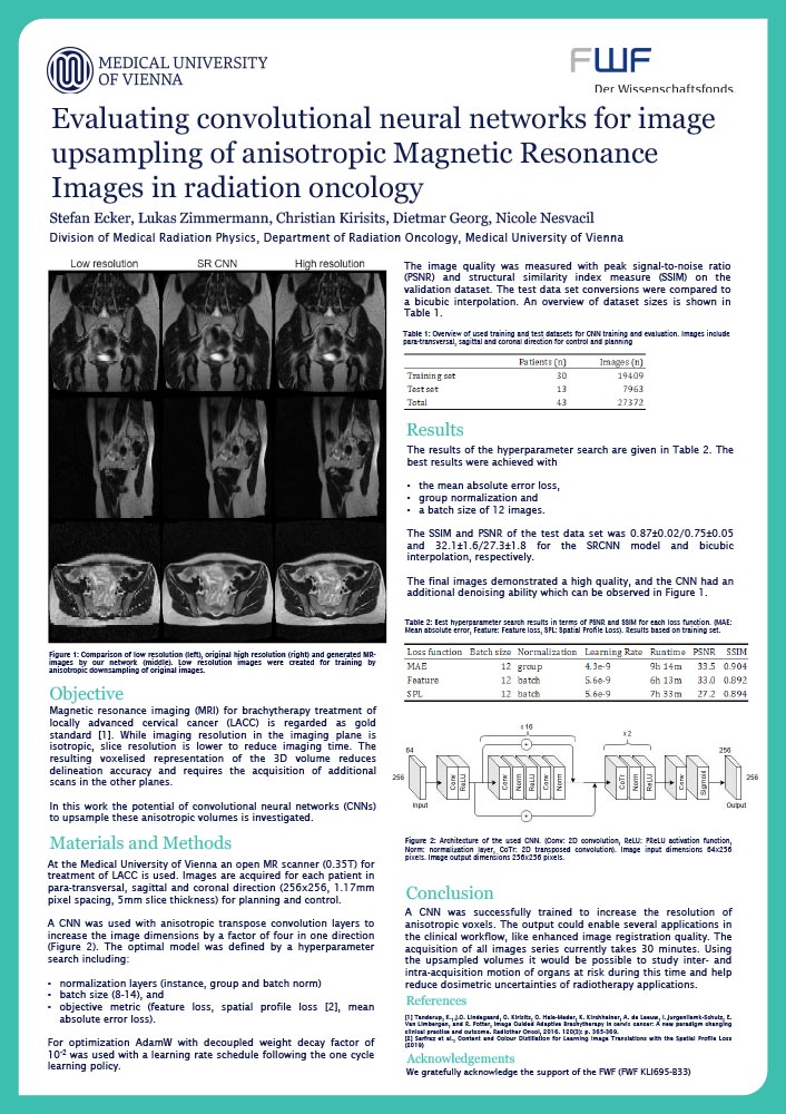
Stefan Ecker
Division of Medical Radiation Physics, Department of Radiation Oncology, Medical University of Vienna
Introduction
Convolutional neural networks (CNN) have shown great success for higher order interpolation image upsampling. In magnetic resonance imaging (MRI) resolution in the imaging plane is isotropic while slice resolution is often lower. At the medical university of Vienna an open MR scanner (0.35T) for brachytherapy treatment of locally advanced cervical cancer (LACC) is used. Images are acquired for each patient in para-transversal, sagittal and coronal direction (256x256, 1.17mm pixel spacing, 5mm slice thickness). In this work the potential of CNNs to upsample these volumes is analysed.
Influence of bone suppression on accuracy and robustness of tumor motion monitoring using intensity-based 2D/3D registration
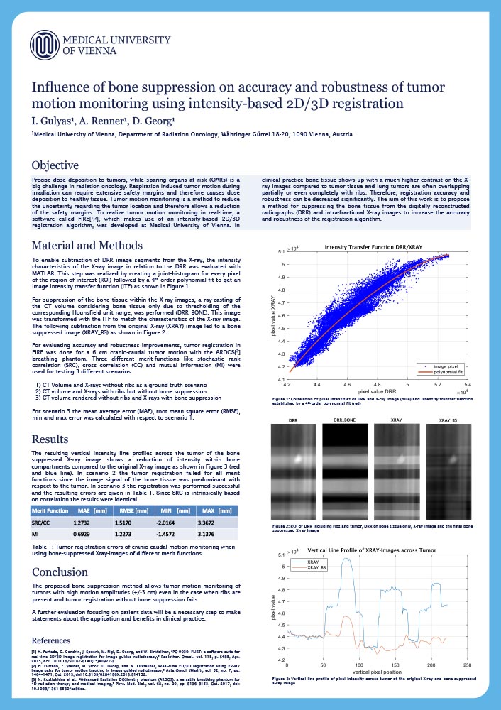
Ingo Gulyas
Department of Radiation Oncology, Medical University of Vienna
Introduction
In radiation therapy of moving lung tumors (breathing) safety margins are introduced to ensure irradiation of the tumor tissue along the internal tumor pathway. Tumor motion monitoring is a method to reduce the uncertainty of the tumor location and the first step towards tumor tracking, which ultimately allows for a reduction of safety margins. Thus, dose deposition in healthy tissue is minimized and side effects in patients are reduced.
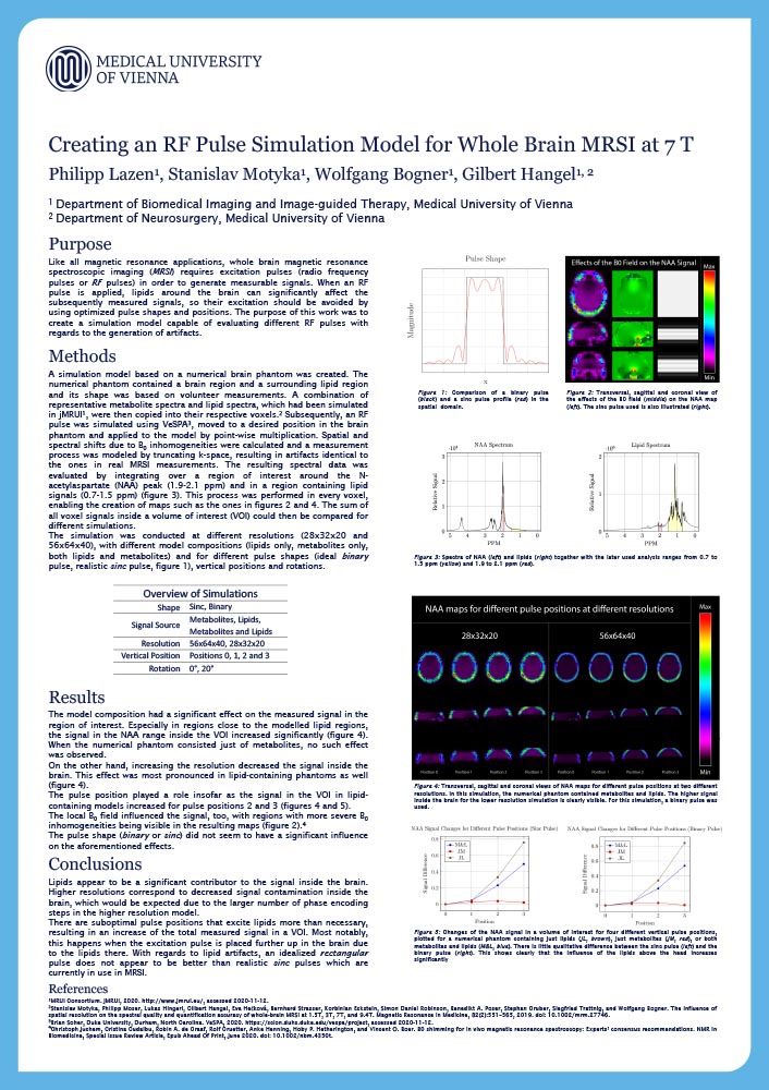
Creating an RF Pulse Simulation Model for Whole Brain MRSI at 7 T
Philipp Lazen
Department of Biomedical Imaging and Image guided Therapy, Medical University of Vienna
Introduction
Like most magnetic resonance applications, whole-brain magnetic resonance spectroscopic imaging generally requires excitation pulses (RF pulses). Lipids around the brain can significantly affect the measured signals so their excitation should be avoided by using optimal pulse shapes and positions. The purpose of this work was to create a means of evaluating and improving different RF pulses.
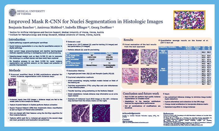
Improved Mask R-CNN for nuclei segmentation in histologic images
Benjamin Bancher
Section for Artificial Intelligence and Decision Support, Medical University of Vienna
Introduction
Accurate identification of cell nuclei is a fundamental step in the analysis of hematoxylin and eosin (H&E)-stained tissue images. Manual nuclei segmentation is a complex and time-consuming procedure prone to intra- and inter-observer variability. Aiding experts through automation of nuclei detection requires robust and efficient computer-based techniques which are currently mainly implemented by deep learning (DL) approaches. Mask R-CNN is a DL architecture proposed to tackle the challenges of instance segmentation. This multi-task method is a combination of several sub convolutional neural networks including a region proposal network, a feature pyramid network and a fully convolutional network that together perform instance segmentation. As such, Mask R-CNN is very well suited for nuclei segmentation through its ability of distinguishing overlapping and touching nuclei, one of the main challenges for current methods. As part of an FFG-funded project exploring DL for nuclei segmentation, a novel pipeline using Mask R-CNN will be explored and introduced. The approach to this challenge is twofold. First, the performance of Mask R-CNN will be assessed on the MoNuSeg2018 dataset. It comprises 30 H&E-stained image patches from seven human organs with a total of 21,623 nuclei for which a direct comparison with 32 other DL based approaches is possible. Following this, the data pool will be expanded with a new dataset of H&E-stained cryo-sections containing 30 images from 10 human organs and 7,596 manually annotated nuclei, the first of its kind. This will grant further insight into the pipeline’s performance and generalization ability. It will also provide the steppingstone for improvement of the current Mask R-CNN pipelines, in particular in regard to the method’s ability to delineate overlapping and touching nuclei. This work will help pathologists’ ability to perform robust and fast analysis of microscopic images through the use of automated nuclei segmentation.
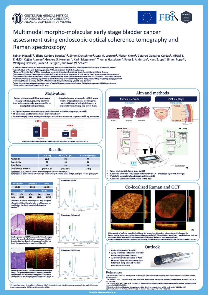
Multimodal morpho-molecular early stage bladder cancer assessment using endoscopic optical coherence tomography and Raman spectroscopy
Fabian Placzek
Center for Medical Physics and Biomedical Engineering, Medical University of Vienna
Introduction
Although the gold standard in bladder cancer assessment - visualization by white light endoscopy – detects 97-100 % of the muscle-invasive bladder cancer, the detection of non-muscle invasive bladder cancer (NMIBC) is still challenging. Different types of NMIBC have a five-year recurrence rate of 20-80 %.
Optical coherence tomography (OCT) and Raman spectroscopy (RS) are promising imaging techniques to assist urologists to detect suspicious areas on site. OCT provides cross-sectional information of tissue with high depth resolution and a penetration depth up to 2 mm, suitable for detection of NMIBC lesions. While OCT gives morphological insight of the tissue, RS reveals the chemical information of the underlying tissue. In combination, OCT and RS complement each other and make morphological and chemical information accessible to the clinicians.
Using forward-looking endoscopic probes a cohort of 116 biopsies were examined. The imaging system enables co-registration of the complementary OCT/RS data. The results are analyzed by texture feature extraction, RS data processing and histopathological work-up for gaining pathological information of the examined samples. The detection accuracy using OCT is 73 %, and the achieved accuracy for determining the cancer grade by RS is 77 %.
Since only one pathological label is assigned to an entire biopsy, with respect to the highest severity observed, it is wasting useful information. This procedure does not identify heterogenous biopsies, which might show healthy and cancerous bladder wall. The co-localized OCT/RS imaging system could lead to a better understanding of the signals originating from the underlying structures. This would enable a localized understanding of present pathologies within examined samples. The results support the use of OCT as a red flag imaging technique and RS as a more specific modality to obtain molecular insights of a lesion. These results on the multimodal approach are paving the way for the development of a probe that will give clinicians the benefit of a complementary onsite technique during patient examination for immediate diagnosis, rather than waiting for a lengthy histopathological work-up.
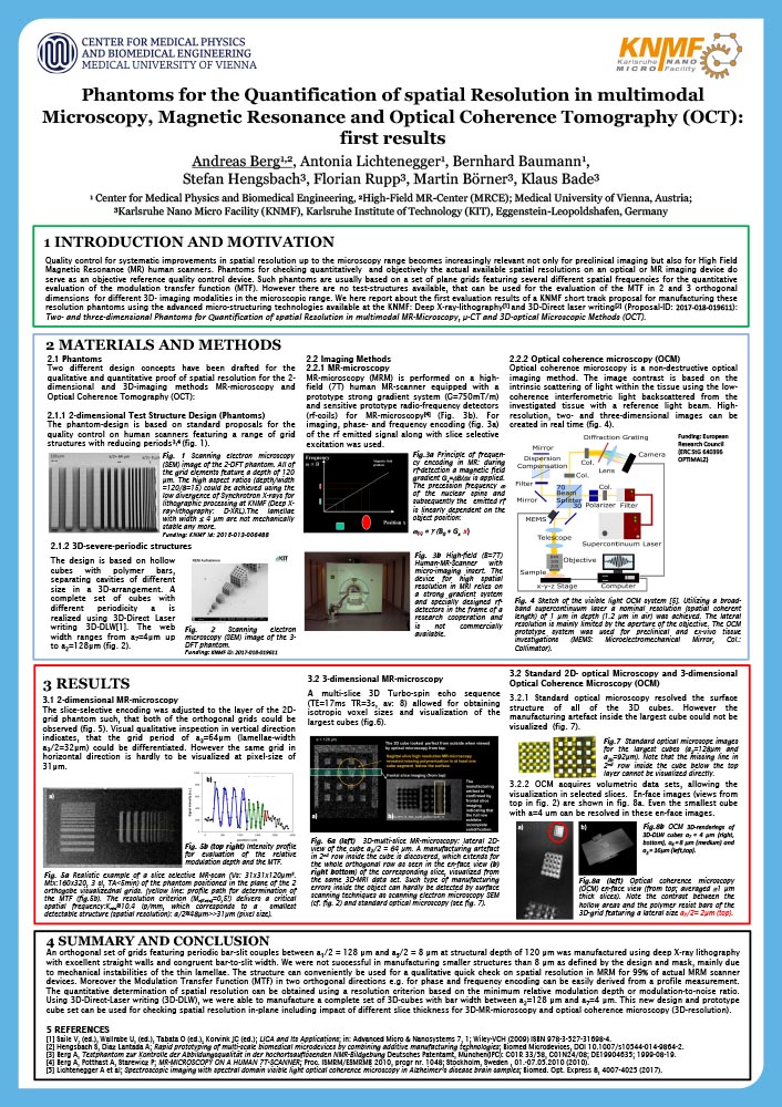
Phantoms for the Quantification of spatial Resolution in multimodal Microscopy, Magnetic Resonance and Optical Coherence Tomography (OCT)
Andreas Berg
Center for Medical Physics and Biomedical Engineering; High-Field MR-Center (MRCE), Medical University of Vienna
Introduction
Quality control for systematic improvements in spatial resolution up to the microscopy range becomes increasingly relevant not only for preclinical imaging and MR-based histology, but also for High Field Magnetic Resonance (MR) human scanners.
For optical microscopy such phantoms are usually based on a set of plane grids featuring several different spatial frequencies for the quantitative evaluation of the modulation transfer function (MTF). However there are no test-structures available, that can be used for the evaluation of the MTF in 2 and 3 orthogonal dimensions for different 3D- imaging modalities as MR-microscopy and OCT in the microscopic range.
The design of the proposed reference resolution standard phantoms as 2D- and 3D test objects comprised orthogonal grids with lateral periods ranging between a1 = 256 µm down to a8 = 2 µm. The demands on miniaturization and spatial accuracy for the establishment of these reference objects are very challenging and can in principle only be fulfilled using the most advanced micro-manufacturing technologies for polymer material as Deep X-Ray Lithography (XRL) and 3D Direct Laser writing (3D-DLW) using 2-photon absorption. We here report about the first evaluation results using a prototype MR-microscopy insert on a High-field human 7T scanner and a prototype OCT microscope on the phantoms manufactured yet within a KNMF proposal (Proposal-ID: 2016-016-014829): Two- and three-dimensional Phantoms for Quantification of spatial resolution in multimodal MR-microscopy, μ-CT and 3D-optical microscopic methods (OCT).
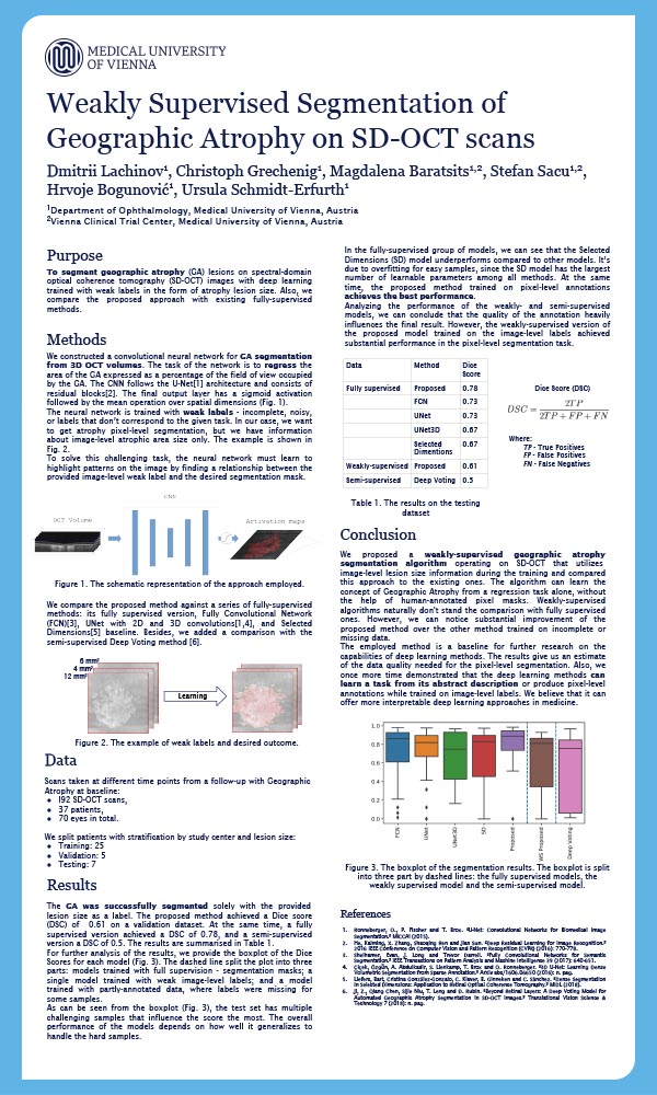
Weakly Supervised Segmentation of Geographic Atrophy on SD-OCT scans
Dmitrii Lachinov
Department of Ophthalmology, Medical University of Vienna
Introduction
To segment geographic atrophy (GA) lesions on spectral-domain optical coherence tomography (SD-OCT) images with deep learning trained with weak labels in the form of atrophy lesion size. In addition, we compare the proposed approach with existing fully pixel-level supervised methods.
We constructed a convolutional neural network for GA segmentation from 3D OCT volumes. The task of the network is to regress the area of the GA expressed as a percentage of the field of view occupied by the GA. The CNN follows the U-Net architecture and consists of residual blocks. The final output layer has a sigmoid activation followed by the mean operation. The neural network learns to highlight patterns on the image by finding a relationship between the provided weak label and the desired segmentation mask. We compare the performance of our method against the supervised version of our algorithm and against the semi-supervised deep-voting model.
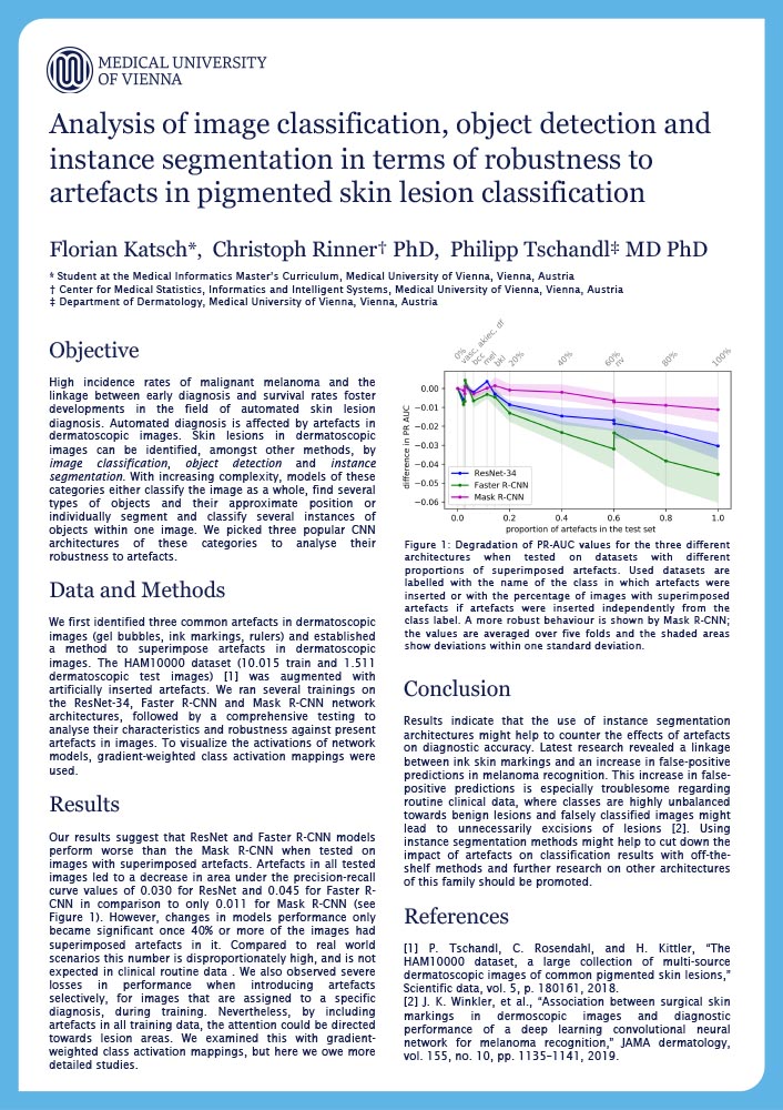
Analysis of image classification, object detection and instance segmentation in terms of robustness to artefacts in pigmented skin lesion classification
Florian Katsch
Student at the Medical Informatics Master’s Curriculum, Medical University of Vienna
Introduction
High incidence rates of malignant melanoma and the linkage between early diagnosis and survival rates foster developments in the field of automated skin lesion diagnosis. Automated diagnosis is affected by artefacts in dermatoscopic images. These artefacts in images can be identified amongst other methods by image classification, object detection and instance segmentation. We picked three popular CNN architectures of these categories to analyse their robustness to artefacts.
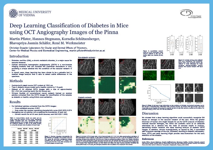
Deep Learning Classification of Diabetes in Mice using Optical Coherence Tomography Angiography Images of the Pinna
Martin Pfister
Christian Doppler Laboratory for Ocular and Dermal Effects of Thiomers
Center for Medical Physics and Biomedical Engineering, Medical University of Vienna
Introduction
Diabetes mellitus (DM), a chronic metabolic disorder, is a major cause for vascular diseases. Optical coherence tomography angiography (OCTA) is a non-invasive imaging modality that can visualize the superficial vasculature in skin, providing a unique window into the condition of the vascular network in vivo. In recent years, deep learning has been shown to be a valuable tool in medical image analysis that is able to detect subtle differences in the specimen.
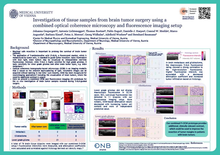
Investigation of tissue samples from brain tumor surgery using a combined optical coherence microscopy and florescence imaging setup
Johanna Gesperger
Center for Medical Physics and Biomedical Engineering, Medical University of Vienna
Division of Neuropathology and Neurochemistry, Department of Neurology, Medical University of Vienna
Introduction
Optical coherence tomography-based microscopy (OCM) is an imaging modality based on the inherent backscattering of light. Volumetric images can be acquired without labeling in real time. Just recently, OCM has been increasingly recognized in various fields such as neuroimaging where the need for additional contrast parameters, i.e. during tumor resection, is urgent. Maximal safe resection is of utmost importance to prolong the survival of brain tumor patients. One particularly important tool to guide surgery is the application of 5-aminolevulinc acid (5-ALA), a fluorescent marker which is metabolized by tumor cells. Upon illumination with blue light, specific brain tumors can be visualized by intraoperative real-time fluorescence (FL). While 5-ALA is highly sensitive for high grade gliomas, it lacks sensitivity for lower grade gliomas and other brain tumor types.
Here, we present a multimodal imaging approach for the ex-vivo investigation of brain tumor samples resected during 5-ALA-guided surgery. Volumetric OCM intensity and FL data were acquired with our prototype, post-processed and evaluated against the gold standard histology. Attenuation (ATT) coefficients, parameters for light scattering and absorption, were calculated along with high-resolution FL intensities. In glioblastoma and brain metastases, the intraoperatively assessed macroscopic 5-ALA FL status showed a strong correlation to the high-resolution FL as measured with our prototype. Higher FL intensity was associated with decreased ATT coefficients and increased tumor infiltration upon histology. Lower grade gliomas, however, did not express any macroscopic FL in 32/34 cases. Still, sub-visual FL could be detected in 11 biopsies with our setup. More notably, ATT values were shown to decrease with increasing grade of malignancy independent of 5-ALA status. Our results show that this prototype provides additional, clinically relevant contrast, which could be used to improve resection of tumor margins.
OCT on a Chip: In vivo three-dimensional Swept Source and Spectral Domain Optical Coherence Tomography and angiography using Photonic Integrated Circuits
Elisabet A. Rank
Center for Medical Physics and Biomedical Engineering. Medical University of Vienna
Introduction
Today, optical coherence tomography (OCT) is considered as a standard imaging technique for ophthalmic care which has massively advanced in terms of resolution as well as contrast in the past few decades. However, comparably little effort has been taken to reduce size and costs of such systems. With a footprint of ~1 m2 and costs of up to 100.000 Euro such a system is both, space and cost wise, a large investment. As these parameters are becoming more and more critical in medical facilities, there is a strong need to lower costs and miniaturize OCT engines.
Photonic integrated circuits (PIC) are an attractive platform to propagate and handle light within a compact area. For OCT applications PICs give the outlook to make systems more robust, smaller and cheaper, which is a pre-requisite for their use in point-of-care diagnostic settings.
We investigated passive photonic building blocks for OCT application. A PIC based interferometer was used for swept source OCT. A fiber couples light from the swept source to the chip, on which a 90/10 coupler split the light into the sample and reference arm. Back-reflected light was interfered on the chip and collected with two photodiodes of a dual balanced detector. A system sensitivity of 96 dB at eye safe laser powers and an axial resolution of 5.5 µm in air were measured.
Furthermore, arrayed waveguide gratings (AWG), the on-chip equivalent to a discrete diffraction grating, with various output channels (256 and 512) were used for spectral domain (SD) OCT. Interfered light from a fiber based SD-OCT system was coupled into the AWG where it was spectrally separated into the output waveguides which are forwarded to the end facet of the chip. The light was then projected onto the individual pixels of a commercial CCD camera. Here a system sensitivity of around 90 dB and axial resolutions range from 11 µm to 7 µm, depending on the used AWG.
The retina of a healthy volunteer was imaged in various areas of the retina using these systems, demonstrating for the very first time that PIC-based OCT has a realistic chance of future commercialization. As a benchmark, the retina of the healthy volunteer was also imaged with a commercial SD-OCT at 840 nm and compared to the on-chip systems.
The Rapid-Fire pitches are taking place during the Science I session at 13:00-13:55.
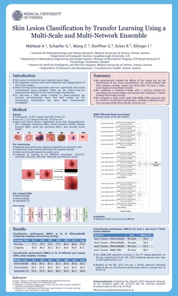
Skin Lesion Classification by Transfer Learning Using a Multi Scale and Multi Network Ensemble
Amirreza Mahbod
Institute of Pathophysiology and Allergy Research, Medical University of Vienna
Introduction
Skin cancer is among the most common cancer types in the white population and consequently computer aided methods for skin lesion classification based on dermoscopic images are of great interest. A promising approach for this uses transfer learning to adapt pre-trained convolutional neural networks (CNNs) for skin lesion diagnosis. Since pre-training commonly occurs with natural images of a fixed image resolution and these training images are usually significantly smaller than dermoscopic images, downsampling or cropping of skin lesion images is required. This however may result in a loss of useful medical information, while the ideal resizing or cropping factor of dermoscopic images for the fine-tuning process remains unknown.
We investigate the effect of image size for skin lesion classification based on pre-trained CNNs and transfer learning. Dermoscopic images from the International Skin Imaging Collaboration (ISIC) skin lesion classification challenge datasets are either resized to or cropped at six different sizes ranging from 224x224 to 450x450. The resulting classification performance of three well established CNNs, namely EfficientNetB0, EfficientNetB1 and SeReNeXt-50 is explored. We also propose and evaluate a multi-scale multi-CNN (MSM-CNN) fusion approach based on a three-level ensemble strategy that utilises the three network architectures trained on cropped dermoscopic images of various scales.
Our results show that image cropping is a better strategy compared to image resizing delivering superior classification performance at all explored image scales. Moreover, fusing the results of all three fine-tuned networks using cropped images at all six scales in the proposed MSM-CNN approach boosts the classification performance compared to a single network or a single image scale. On the ISIC 2018 skin lesion classification challenge test set, our MSM-CNN algorithm yields a balanced multi-class accuracy of 86.2% making it the currently second ranked algorithm on the live leaderboard.
We confirm that the image size has an effect on skin lesion classification performance when employing transfer learning of CNNs. We also show that image cropping results in better performance compared to image resizing. Finally, a straightforward ensembling approach that fuses the results from images cropped at six scales and three fine-tuned CNNs is shown to lead to the best classification performance.
Forward viewing photoacoustic laparoscope using all optical ultrasound detection, flexible fiber bundles and fiber core targeted fast scanning
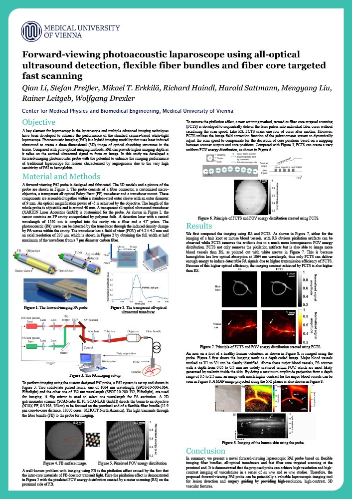
Qian Li
Center for Medical Physics and Biomedical Engineering, Medical University of Vienna
Introduction
A key element for laparoscopy is the laparoscope and multiple advanced imaging techniques have been developed to enhance the performance of the standard camera based white light laparoscope Photoacoustic imaging ( is a hybrid imaging modality that uses laser induced ultrasound to create a three dimensional 3 D) image of optical absorbing structures in the tissue Compared with pure optical imaging methods, PAI can provide higher imaging depth as it relies on the excited ultrasound signal to form an image In this study we developed a forward imaging photoacoustic probe with the potential to enhance the imaging performance of traditional laparoscope for lesions characterized by angiogenesis due to the very high sensitivity of PAI to hemoglobin.
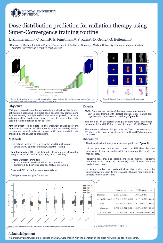
Dose distribution prediction for radiation therapy using Super-Convergence training routine
Lukas Zimmermann
Department of Radiation Oncology, Medical University of Vienna
Introduction
With state-of-the-art radiation therapy techniques, treatment planning and the close distribution optimization according to clinical goals became very complex and time consuming. Multiple techniques were proposed to perform automatic dose prediction but due to inconsistent data sets a direct comparison is not possible.
Influence of delineation modality on overall accuracy of machine learning predictive model on colorectal carcinoma
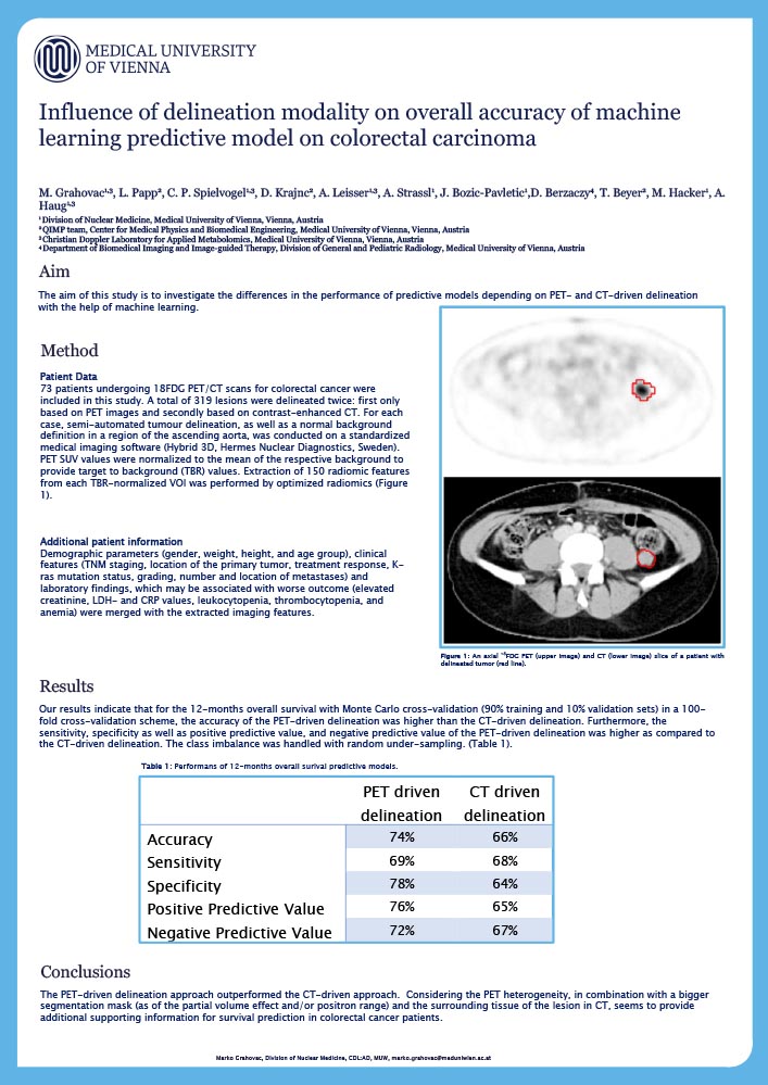
Marko Grahovac
Division of Nuclear Medicine, Medical University of Vienna
Christian Doppler Laboratory for Applied Metabolomics
Introduction
Positron Emission Tomography (PET) / Computer Tomography (CT) support diagnosis and follow-up of patients with colorectal cancer (CRC). To date, the association of the PET and CT-driven in vivo radiomics features with the overall survival (OS) is not well explored. The aim of this study was to investigate the differences in the performance of predictive models depending of PET- and CT-driven delineation with the help of machine learning.
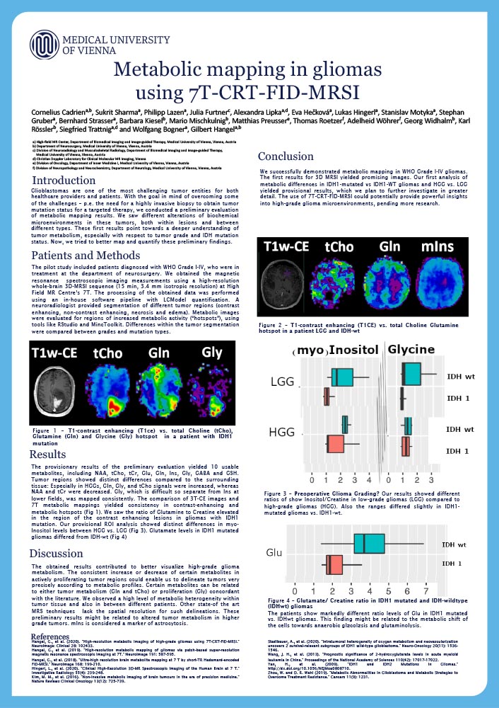
Metabolic mapping in gliomas using 7T-CRT-FID- MRSI
Cornelius Cadriena
High-field MR Center, Department of Biomedical Imaging and Image-guided Therapy
Department of Neurosurgery, Medical University of Vienna
Introduction
Glioblastomas create altered biochemical microenvironments to grow and thrive. Using recent advances in magnetic resonance metabolic imaging, we mapped heterogenous metabolic behavior with 7T-CRT-FID-MRSI.
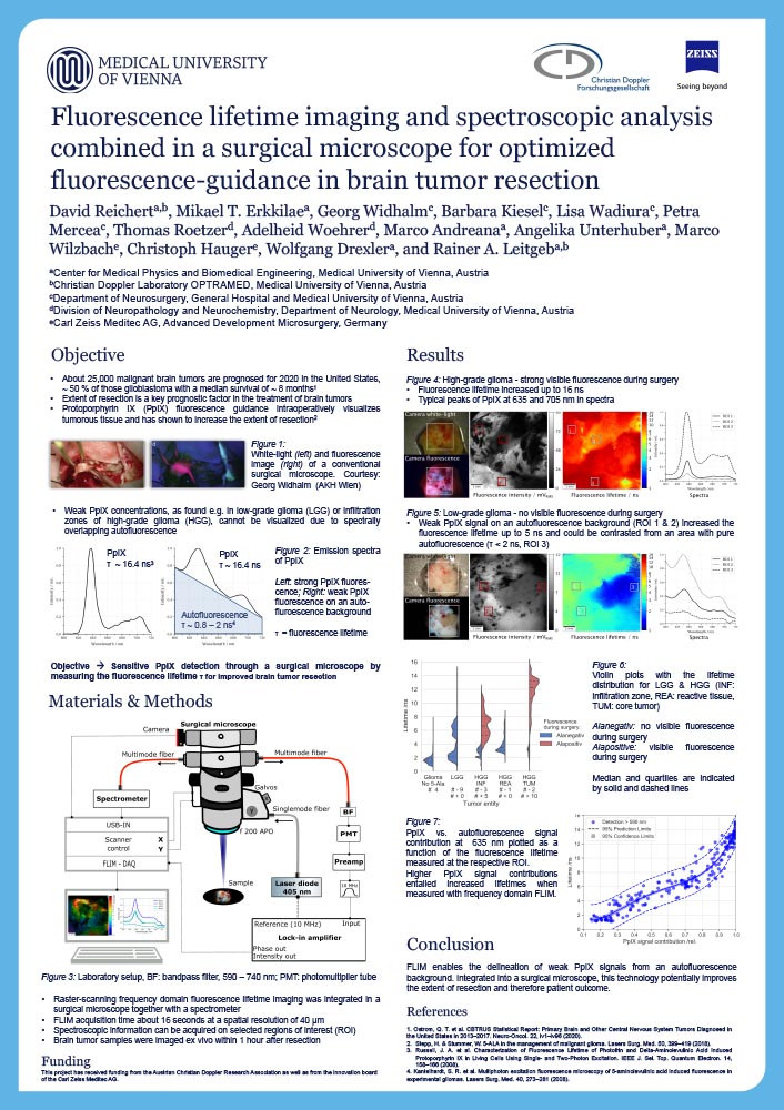
Fluorescence lifetime imaging and spectroscopic analysis combined in a surgical microscope for optimized fluorescence-guidance in brain tumor resection
David Reichert
Center for Medical Physics and Biomedical Engineering, Medical University of Vienna
Christian Doppler Laboratory OPTRAMED
Introduction
Protoporphyrin IX (PpIX) fluorescence-guided surgery has established as a standard for enhanced resection of malign glioma. However, low-grade glioma (LGG) or weakly infiltrated brain parenchyma often emit weak PpIX fluorescence and are hard to distinguish from non-pathological tissue with conventional surgical microscopes. What is more, low PpIX concentrations compete with spectrally overlapping tissue autofluorescence, inherently limiting the sensitivity of fluorescence intensity based PpIX detection.