by Gilbert Hangel
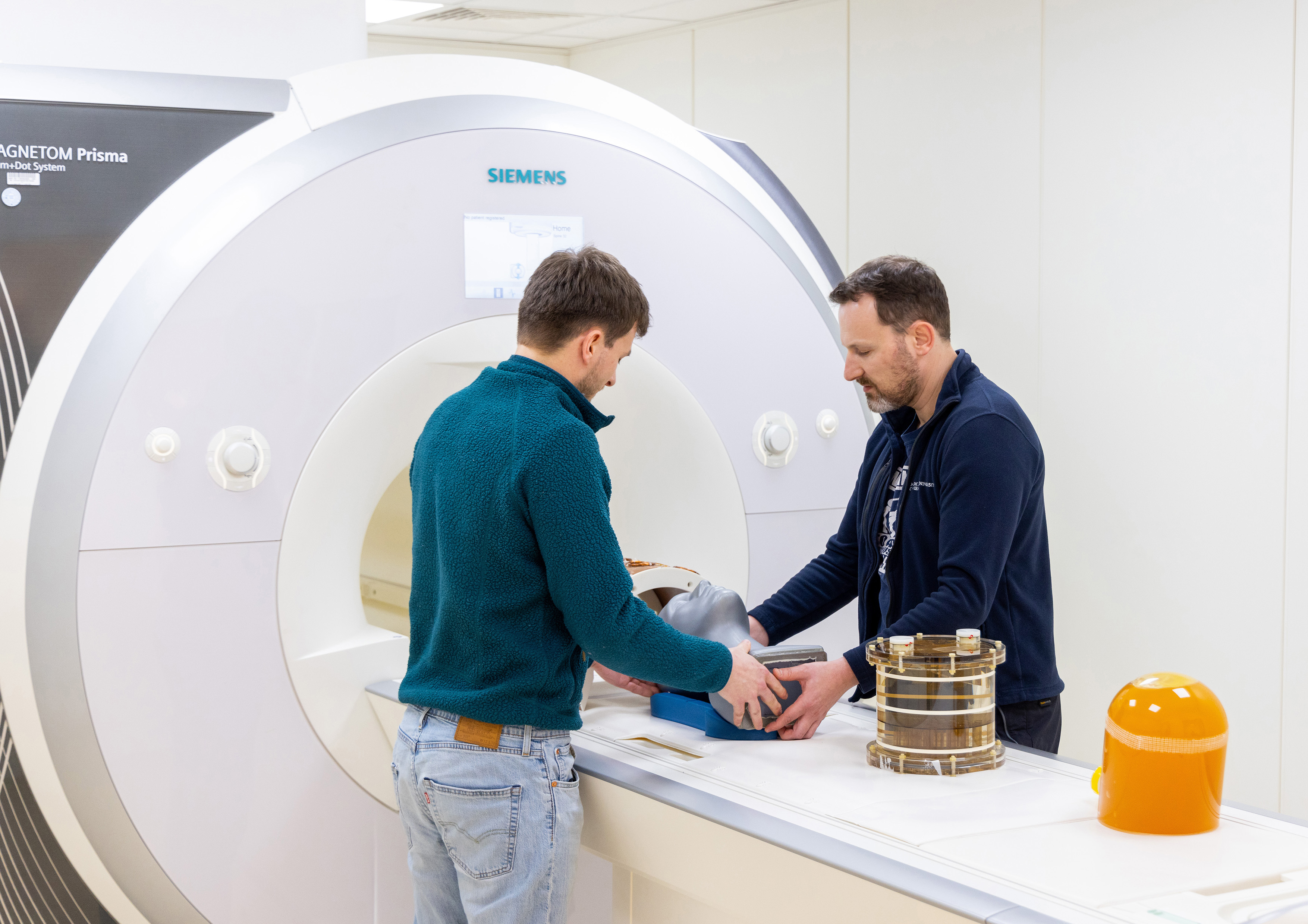
Photos by: MedUni Wien/feelimage / Wolfgang Bogner (Image 1) and Gilbert Hangel (Portrait)

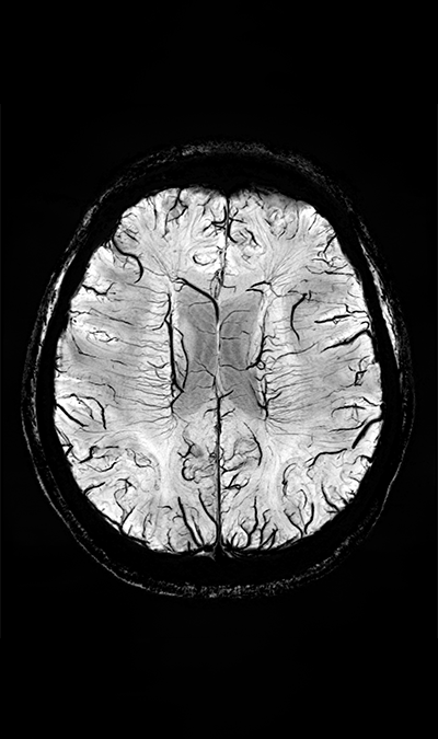
Magnetic resonance imaging (MRI) is one of the most important clinical imaging modalities available to date. Using this technology, we can measure the amount of water and electromagnetic properties in biological tissues, which allows us to make images that show a contrast between these different tissues. Depending on the measurement settings, we can make different structures or changes in the human body visible. Routine MRI protocols can image the structure of the human brain with great detail, but other possibilities such as function and neurochemistry are far less developed.
At the High Field MR Centre of the Medical University of Vienna in the heart of Europe, we work to not only make images of the human brain's structure, but we also want to learn more about the metabolism and neurochemistry that define the healthy and diseased human brain. Neurochemistry is the study of the chemical processes that occur in the nervous system, including the functions of neurotransmitters, neuropeptides and other molecules involved in neurological activity.
To this end we use proton magnetic resonance spectroscopic imaging (MRSI), with which we do not measure signals from water molecules as in MRI, but from other molecules found in the human body. Not all biochemicals can be measured, but the ones imageable include the neurotransmitter glutamate or the metabolite creatine. As the concentrations of these target molecules in our bodies are far lower than that of water, the available signal-to-noise ratio (SNR), which determines the balance between scan time and image resolution, is much lower as well.

"At the High Field MR Centre, we can use Austria’s only 7 Tesla MRI scanner for this task. Worldwide, there are only around 100 similar machines."
Clear image of 5-10 Molecules in the human brain in only 15Min
At the High Field MR Centre, we can use Austria’s only 7 Tesla MRI scanner for this task. Worldwide, there are only around 100 similar machines. Its magnetic field is much stronger than that of the MRIs used in clinics, which practically means much higher SNR and allows us to differentiate more molecules in the brain. Using the 7T MRI allows us to introduce a new MRSI method that allows faster scans and higher resolutions than previous technologies.
With this world-leading technology, we can simultaneously image around 5-10 different molecules in the human brain with a resolution of 3.4 mm in 15 minutes.
In first studies, we have identified promising applications in a range of neuropathologies like gliomas or multiple sclerosis. Gliomas are a type of brain tumour that originate from glial cells, which are cells that provide protection and support for neurons in the brain. By measuring the levels of different molecules, such as neurotransmitter glutamate or the metabolite creatine, we can learn more about the underlying mechanism of glioma development and progression. This can help with earlier diagnosis, as well as development of more personalized, targeted and effective treatments. Multiple sclerosis (MS) is a neurological disorder where the immune system mistakenly attacks the myelin sheath, a protective covering that surrounds nerve fibres, causing an information disruption between the brain and the rest of the body. MRSI allows us to get valuable insights to learn more about the changes that occur in the brain during the progression of the disease, which may lead to the identification of new markers and development of new and more target treatments and drug development.

Our research group, led by Prof. Wolfgang Bogner, the recent recipient of an ERC consolidator grant, is not only equipped with modern MRI scanners, but also benefits from the environment of one of Europe’s largest hospitals to amplify clinical research. Located near the city centre, our team also enjoys Vienna’s famous high quality of living, be it Vienna’s many cultural offerings or the easy access to parks and nature.
Currently, our research efforts concentrate onto improvements in technology, such as faster image reconstruction, motion correction or artefact reduction, to progress towards implementation in clinical MRI systems on the one hand and the investigation of applications in pathologies in collaboration with many clinical departments, e.g., Radiology, Neurosurgery, and Neurology. Current PhD projects within these topics include the correlation of oncometabolites to clinical classifications and molecular markers, the identification of neurochemical changes in diseases before structural changes can be detected, or the improved stability of molecular quantification.
If you are interested in our research, have a background in physics, medicine, biomedical engineering or related fields, and want to pursue a PhD, do not hesitate to contact us via one of the Medical University’s PhD Calls.