
Hoch-ortsauflösende Bildgebung bis in den Mikroskopiebereich auf einem Human MR-scanner
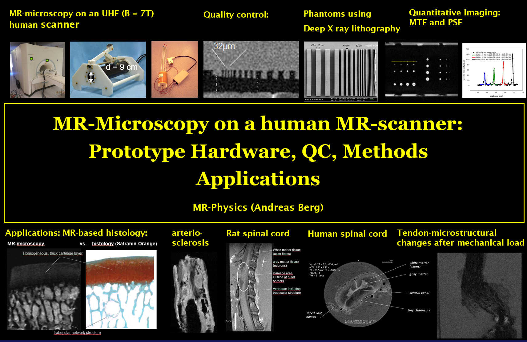
Die räumliche Auflösung ist neben dem Kontrast das wichtigste Qualitätskriterium für die diagnostische Bildgebung.
Auf klinischen MR-Scannern können typischerweise strukturelle Änderungen im mm-Bereich aufgelöst werden. Die kleinsten Voxel-Größen als kleinstes informationstragendes Bildelement liegen selbst bei UHF MR-Scannern bei nur ca. (0.4 mm)3.
Unsere zentralen Forschungsfragen sind:
- Welche Ortsauflösungen sind auf einem Humanscanner mit Zusatzausstattung möglich?
- Welche Faktoren begrenzen die Ortsauflösung und wie kann sie probenspezifisch verbessert werden?
- Wie stellen sich pathologische Veränderungen MR-mikroskopisch im Gewebe dar?
- Kann eine MR-basierte Histologie an entnommenem Gewebe wertvolle Informationen für schnelle Diagnosen liefern, ohne die Proben für weitere Untersuchungen unbrauchbar zu machen?
- Können mit speziellen MR-Messverfahren auch Gewebe und Implantate strukturell dargestellt werden, die mit Standard-Methoden nicht dargestellt werden können (z.B. Sehnen, Zähne, Polymere)?
- Welchen Beitrag kann die MR-gestützte hoch-ortsauflösende 3D Polymergel-Dosimetrie für die Validierung von Bestrahlungsplänen in der Strahlentherapie leisten?

Andreas Berg
GruppenleiterORCID: 0000-0002-2190-5242
T +43 1 40400-37730
andreas.berg@meduniwien.ac.at
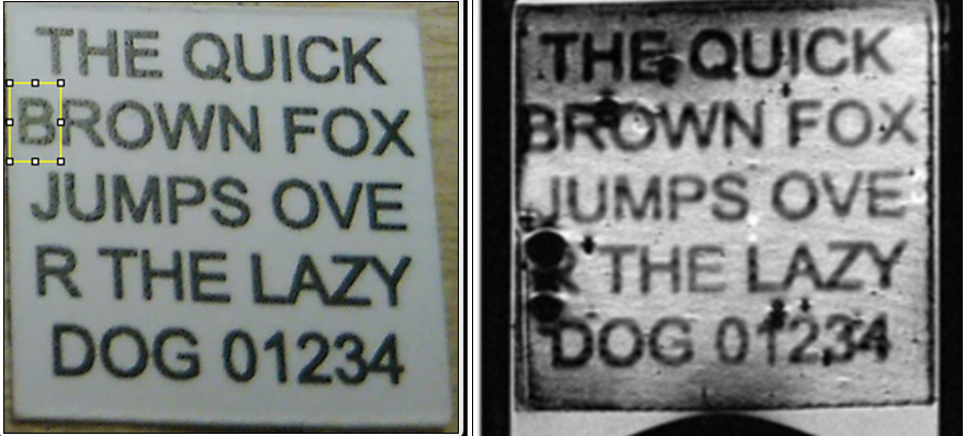
3D-MR-Mikroskopie mit isotropen Voxeln bietet bei derzeit beschränkten Probengrössen (ca. 40 mm x 19 mm x 19 mm) die Perspektive für nicht-zerstörendes Lesen wertvoller Handschriften oder Drucken in Büchern oder Papier-Rollen). Die erreichten minimalen Voxelgrössen von (20 µm)3 sind auf einem Humanscanner einzigartig.
Beteiligte Forschungsgruppen
Publikationen
- Visualization of text on bowed sheets via High-resolution 3D-Magnetic Resonance Micro-imaging for potential reading of closed books: the proof-of-concept (Nature Communcations Engineering, 2026)
Body >> Muskuloskeletal
MR-basierte Histologie
UTE Bildgebung der Sehne
Hardware >> Phantome
Auflösungsphantome für MR-Mikroskopie
Entwicklung von MR-sichtbaren Polymeren
- Multi-Center CORREL µ-imaging of a tendinopathy model). KNMF Projektnr. 2025-033-032280.
- Multi-Purpose-Phantoms for the Quality Control in Multimodal Slice-selective MR-Microscopy, μ-CT and slice- selective optical Microscopy (OCT). KNMF Projektnr. 2021-025-030114 (finished 2026).
- Magnetic resonance –Broadline-imaging. Hochschuljubiläumsfonds der Stadt Wien; Projektnr.: H-247/2001 (finished 2025).
- MR-based dosimetry of High-LET radiation" (3D )proton and neutron dosimetry . Hochschuljubiläumsfonds (HSJ) der Stadt Wien, Projectnr.: H-1208/2003 (finished 2025).
- Two- and three-dimensional Phantoms for Quantification of spatial Resolution in multimodal MR-Microscopy, μ-CT and 3D-optical Microscopic Methods (OCT). KNMF Projektnr. 2017-018-019611 (finished 2023).
- MR visible polymer for medical imaging applications. Austria Wirtschafts-Service (AWS) mit Mitteln aus der Nationalstiftung für Forschung, Technologie und EntwicklungAWS P2372591 (finished 2023),
- Anisotropic Phantoms for quantitative Tests on the Limits in spatial Resolution in MRI using PSF and Diffusion Anisotropy. KIT Projektnr. 2015-015-009869 (finished 2019).
- 2-3-Dimensional μ-Structures for Magnetic Resonance Microscopy. KIT Projektnr. 2015-013-006488 (finished 2017).
- Multi-parametric visualization of knee joint ultra-structure. FWF Projektnr.: I690.
- Magnetic resonance (MR-) based Polymer-Gel-Dosimetry for applications in precision therapy (Brachytherapy, IMRT, stereotactic irradiation). Österreichische Nationalbank Wissenschaftsfonds.
- Dosimetry of highly selective therapeutic radiation procedures using parameter selective MR-microscopy. Hochschuljubiläumsfonds der Stadt Wien HSJ Wien H194/99.
- NMR-Microscopy for the early detection of diabetic Neuropathy". FWF Project: P12041, 1996.
- Visualization of text on bowed sheets via High-resolution 3D-Magnetic Resonance Micro-imaging for potential reading of closed books: the proof-of-concept (Nature Communications Engineering, 2026)
- A phantom for the quantitative determination and improvement of the spatial resolution in slice-selective 2D-FT magnetic resonance micro-imaging and -microscopy based on Deep X-ray Lithography (DXRL) (Front. Phys., 2023)
- Transverse Relaxation Anisotropy of the Achilles and Patellar Tendon Studied by MR Microscopy (J Magn Reson Imaging, 2022)
- Basic Properties of a New Polymer Gel for 3D-Dosimetry at High Dose-Rates Typical for FFF Irradiation Based on Dithiothreitol and Methacrylic Acid (MAGADIT): Sensitivity, Range, Reproducibility, Accuracy, Dose Rate Effect and Impact of Oxygen Scavenger (Polymers, 2019)
- MR-µ-imaging based 3-dimensional-polymer gel dosimetry in comparison to 2D-film and 1D-diamond dosimetry of mm-sized photon pencil beams (Biomedical Engineering-Biomedizinische Technik, 2017)
- Diffusivity- and T2-imaging at 3 Tesla for the Detection of Degenerative Changes in Human-Excised Tissue with High Resolution: Atherosclerotic Arteries (Investigative Radiology, 2003)
- METHOD FOR PRODUCING A LIGHT-CURABLE RESIN COMPOSITION (EPT-Application, 2022)
- Andreas Weiland (FH Campus Wien, Bsc. 26.05.2024); Parameter selective Magnetic Resonance Imaging of micro-capsule modified polymers manufactured for MR-visibility.
- Paul Perry (TU-Wien, Diplomarbeit, 01.09.2023); High-field Magnetic Resonance-based visualization of new polymer and resin materials for tissue mimicking phantoms.
- Katharina Buchner (TU Wien, Atominstitut, Dipl. Ing., 26.01.2022); The impact of high Z elements as add-ons to gel dosimeters for increasing local dose in radiation therapy.
- Muzafar Khan (Medizinische Universität Wien, PhD, 08/2018); Magnetic Resonance Imaging based polymer gel dosimetry for radiation therapy: basic properties of new normoxic polymer gels for high dose rates.
- Markus Ziegner (ULG Medical Physics Medical Univ. Vienna, Abschlussarbeit, 2016); MR-based polymer gel dosimetry on MAGIC type polymer gels: neutron dosimetry.
- Fabian Valka (Univ. Vienna, Master, 25.11.2015); MRI Microscopy on Biocompatible Materials and Polymers with Short T2* Times: Validation and Quality Control of UTE Pulse Sequences for Short Detection Times on a Microscopy Insert to a High-Field Human MR Scanner.
- Dr. Karin Wiesauer (Medical Univ. Vienna, Postgradualer Universitätslehrgang/ULG Medical Physics, Abschlussarbeit, 08/2012); MR Microscopy at 7T using the UTE pulse sequence with special focus on imaging of dental composites.
- Christian Horn (TU Wien, master, 2012); Parameter selective MR-microimaging of Biomaterials with short T2.
- Christian Bayreder ( Dr. , Medizin. Universität Wien, 05/2008,) High resolution Magnetic Resonance based dosimetry using nomoxic polymer gels.
- Markus Pfleger (Techn, Univ. Vienna., master, 19.08.2007); Magnetresonanz-Breitlinienbildgebung an halbfestem biologischem Gewebe, Polymeren und Dental-Compositen.
- Peter Jungbauer (Univ. Vienna, master, 2001); Hochortsauflösende Hochfeld-Magnetresonanz-Bildgebung and halbfesten Biologischen Materialien.
- Thomas Singer (Techn. Univ. Wien, master, 2001): Diffusivität in einem arthritisch geschädigtem Knopel-Modell.