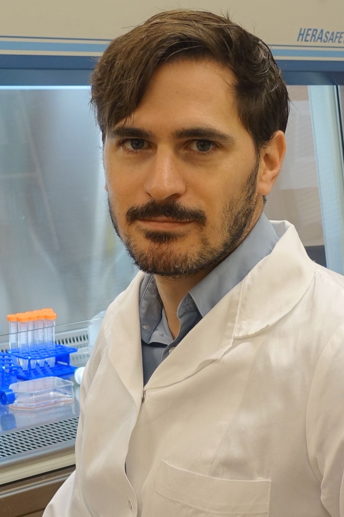
DI Dr. Karl Heinrich Schneider

MedUni Wien RESEARCHER OF THE MONTH May 2019
Acellular vascular matrix grafts from human placenta chorion: Impact of ECM preservation on graft characteristics, protein composition and in vivo performance
Schneider KH, Enayati M, Grasl C, Walter I, Budinsky L, Zebic G, Kaun C, Wagner A, Kratochwill K, Redl H, Teuschl AH, Podesser BK, Bergmeister H.
Biomaterials. 2018 Sep;177:14-26. doi: 10.1016/j.biomaterials.2018.05.045. Epub 2018 May 29.
Abstract:
Small diameter vascular grafts from human placenta, decellularized with either Triton X-100 (Triton) or SDS and crosslinked with heparin were constructed and characterized. Graft biochemical properties, residual DNA, and protein composition were evaluated to compare the effect of the two detergents on graft matrix composition and structural alterations. Biocompatibility was tested in vitro by culturing the grafts with primary human macrophages and in vivo by subcutaneous implantation of graft conduits (n=7 per group) into the flanks of nude rats. Subsequently, graft performance was evaluated using an aortic implantation model in Sprague Dawley rats (one month, n=14). In situ graft imaging was performed using MRI angiography. Retrieved specimens were analyzed by electromyography, scanning electron microscopy, histology and immunohistochemistry to evaluate cell migration and the degree of functional tissue remodeling.
Both decellularization methods resulted in grafts of excellent biocompatibility in vitro and in vivo, with low immunogenic potential. Proteomic data revealed removal of cytoplasmic proteins with relative enrichment of ECM proteins in decelluarized specimens of both groups. Noteworthy, 16 proteins were exclusively preserved in Triton decellularized specimens in comparison to SDS-treated specimens. Aortic grafts showed high patency rates, no signs of thrombus formation, aneurysms or rupture. Conduits of both groups revealed tissue-specific cell migration indicative of functional remodeling.
This study strongly suggests that decellularized allogenic grafts from the human placenta have the potential to be used as vascular replacement materials. Both detergents produced grafts with low residual immunogenicity and appropriate mechanical properties. Observed differences in graft characteristics due to preservation method had no impact on successful in vivo performance in the rodent model.
KEYWORDS: decellularized matrix, human placenta, small diameter vascular grafts, cell migration, biocompatibility,
Selected Literature
1. Schneider KH, Enayati M, Grasl C, Walter I, Budinsky L, Zebic G, et al. Acellular vascular matrix grafts from human placenta chorion: Impact of ECM preservation on graft characteristics, protein composition and in vivo performance. Biomaterials. 2018;177:14-26.
2. Inglis S, Schneider KH, Kanczler JM, Redl H, Oreffo ROC. Harnessing Human Decellularized Blood Vessel Matrices and Cellular Construct Implants to Promote Bone Healing in an Ex Vivo Organotypic Bone Defect Model. Adv Healthc Mater. 2018:e1800088.
3. Hackethal J, Muhleder S, Hofer A, Schneider KH, Pruller J, Hennerbichler S, et al. An Effective Method of Atelocollagen Type 1/3 Isolation from Human Placenta and Its In Vitro Characterization in Two-Dimesional and Three-Dimensional Cell Culture Applications. Tissue Eng Part C Methods. 2017;23(5):274-85.
4. Schneider KH, Aigner P, Holnthoner W, Monforte X, Nurnberger S, Runzler D, et al. Decellularized human placenta chorion matrix as a favorable source of small-diameter vascular grafts. Acta Biomater. 2016;29:125-34.
5. Rohringer S, Hofbauer P, Schneider KH, Husa AM, Feichtinger G, Peterbauer-Scherb A, et al. Mechanisms of vasculogenesis in 3D fibrin matrices mediated by the interaction of adipose-derived stem cells and endothelial cells. Angiogenesis. 2014;17(4):921-33.